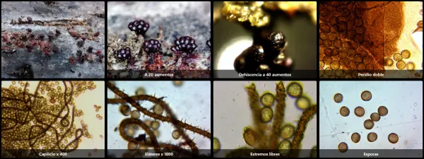
Metatrichia vesparia
Taxonomy
Main synonym
Common names
Not available
Geographic references:
Interactive map with data from GBIF.
Morphological Features:
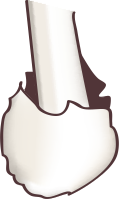
Sporophore developing individually; although fruitbodies of multiple individuals are commonly found on the same substrate, they develop independently. The upper portion consists of several clustered heads (capitula), dark in color and almost black when young. Upon maturity, they rupture to release spores, leaving behind reddish-brown cavities throughout, with an appearance reminiscent of a honeycomb. The size is merely a few millimeters. Accompanying the spores and contributing to their dispersal are a series of elongated sterile fibers that constitute the so-called capillitium. In species of this genus, these filaments are elongated and interwoven, as well as more or less spinose.
Spores spherical, brown to reddish-brown in color, measuring between 8 and 11 μm in diameter, with a probable presence of small warts or short spines.
Stipe formed from the hypothallus mucilage, crimson in color, sometimes poorly defined or frayed in appearance, and generally shorter than the capillitium.
Habitat:
Develops on decaying broadleaf wood in humid areas. The studied specimens were found in February on a decaying beech log (Fagus sylvatica) in the province of Álava.
Observations:
It is a curious and striking species of Myxomycetes, belonging to the Trichia group, characterized by possessing a true capillitium composed of spinose and interwoven fibers, as well as its Myxogaster-like development. It is distinguishable from other species of the genus Trichia, such as Trichia decipiens, by the multiple heads it develops arising from a single stipe.