Clavulinopsis luteoalba
Taxonomy
Main synonym
Common names
Not available
Geographic references:
Interactive map with data from GBIF.
Morphological Features:
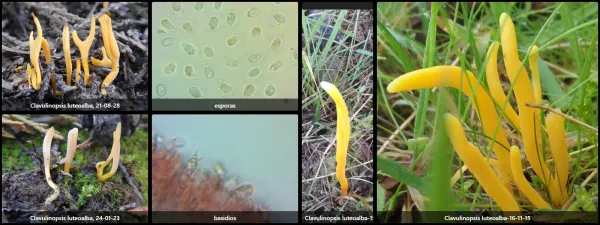
Basidiocarp initially thread-like in shape, it thickens somewhat as it develops, particularly in its central region, resulting in a club-shaped or flattened asparagus-like form. It is uniform, meaning no differentiated parts are discernible. Its color is bright orange, especially in young specimens, while fully developed individuals may exhibit a duller, even yellowish hue. It is small-sized; the specimens found did not exceed 4 cm in height. It is sometimes compressed centrally, but this is a character we have observed to be erratic throughout the family Clavariaceae. In mature specimens, a whitish apical zone is frequently observed, a distinctive character of the species, though not always present. This apex is more acute in young specimens but soon becomes rounded.
Hymenium immersed in the pileipellis of the basidiocarp. The base is narrower and sterile.
Flesh scant and unremarkable, yellowish-orange in color, without distinctive odor or taste.
Habitat:
An eminently heliophilous species that fruits during autumn in grassy meadows and herbaceous clearings within forests. A rare and poorly documented species.
Observations:
This is a small species whose definitive identification requires proper microscopic analysis. Macroscopically similar to Clavulinopsis helvola, from which it is distinguished by having verrucose spores. It can be more readily distinguished from Clavulinopsis fusiformis, which grows in clusters and has bitter flesh. Another related species is Clavulinopsis laeticolor, which features ellipsoid spores with a distinct lateral apiculus.