


Macrocystidia cucumis
(Pers.) Joss.
Study description
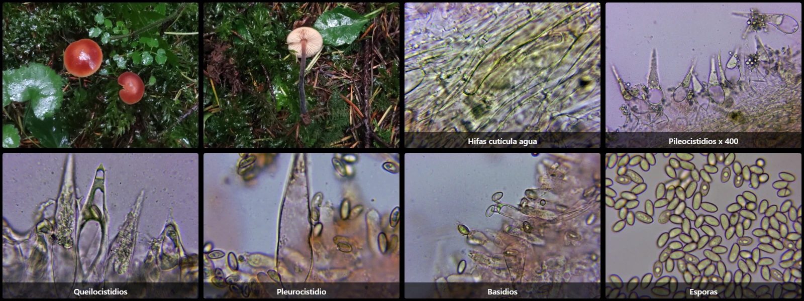
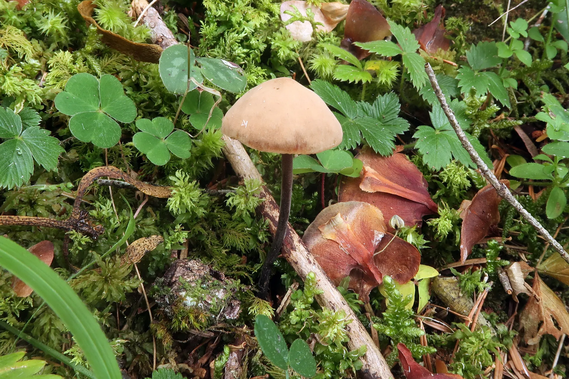
General: Small stature agaric with a reddish-brown pileus and yellowish margin, stipe concolorous darkening to blackish at the base, fishy/cucumber-like odor, characterized by immense cystidia throughout.
Pileipellis: Cutis composed of cylindrical interwoven hyphae 2-6 µm wide with abundant brown intracellular pigment, septate with clamp connections.
Pileocystidia: Emergent, lageniform, ventricose, 65-90 x 16-20 µm.
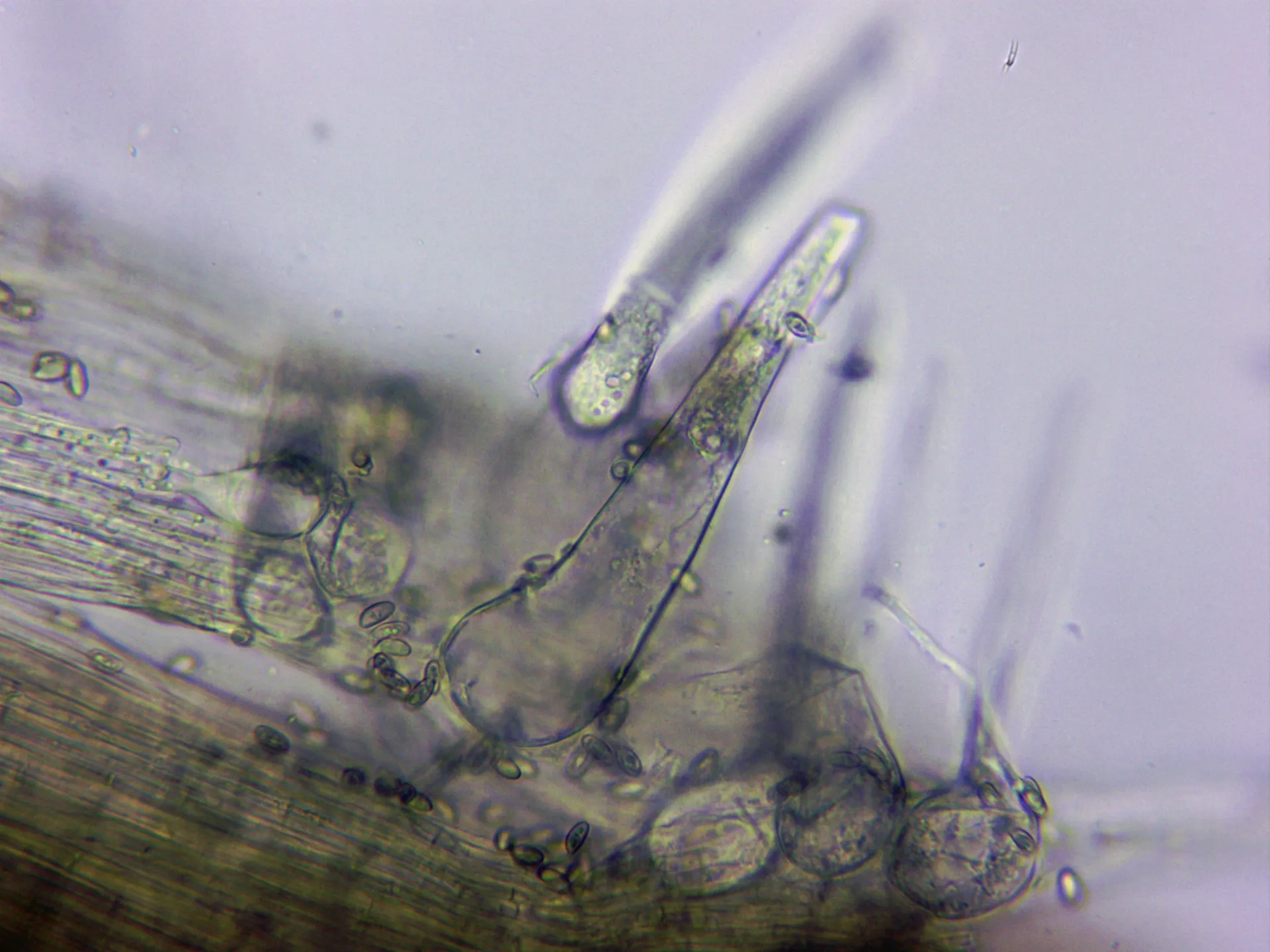
Caulocystidia: Abundant, morphologically similar to pileocystidia.
Cheilocystidia: Fusiform to lageniform, 60-95 x 15-22 µm.
Pleurocystidia: Identical in morphology to cheilocystidia, larger, 70-120 µm long.
Basidia: Cylindrical to slightly clavate, 25-30 x 6-8 µm, with basal clamp connections, 4-spored.
Spores: Cylindric-ellipsoid, smooth, hyaline, moderately thick-walled, 6.5-9 x 3.5-4.5 µm, with an inconspicuous apiculus, containing oil guttules, inamyloid in Melzer's reagent.
Pileipellis: Cutis composed of cylindrical interwoven hyphae 2-6 µm wide with abundant brown intracellular pigment, septate with clamp connections.
Pileocystidia: Emergent, lageniform, ventricose, 65-90 x 16-20 µm.
Caulocystidia: Abundant, morphologically similar to pileocystidia.
Cheilocystidia: Fusiform to lageniform, 60-95 x 15-22 µm.
Pleurocystidia: Identical in morphology to cheilocystidia, larger, 70-120 µm long.
Basidia: Cylindrical to slightly clavate, 25-30 x 6-8 µm, with basal clamp connections, 4-spored.
Spores: Cylindric-ellipsoid, smooth, hyaline, moderately thick-walled, 6.5-9 x 3.5-4.5 µm, with an inconspicuous apiculus, containing oil guttules, inamyloid in Melzer's reagent.
Geographic references
Map generated from global occurrence data from GBIF. GBIF.
Study measurements
Values in Microns (µm)Macroscopic gallery
1 / 6Macrocystidia cucumis - 1 - IMG_0398
Microscopic gallery
1 / 20Macrocystidia cucumis - 10 - Caulocistidios agua x 400