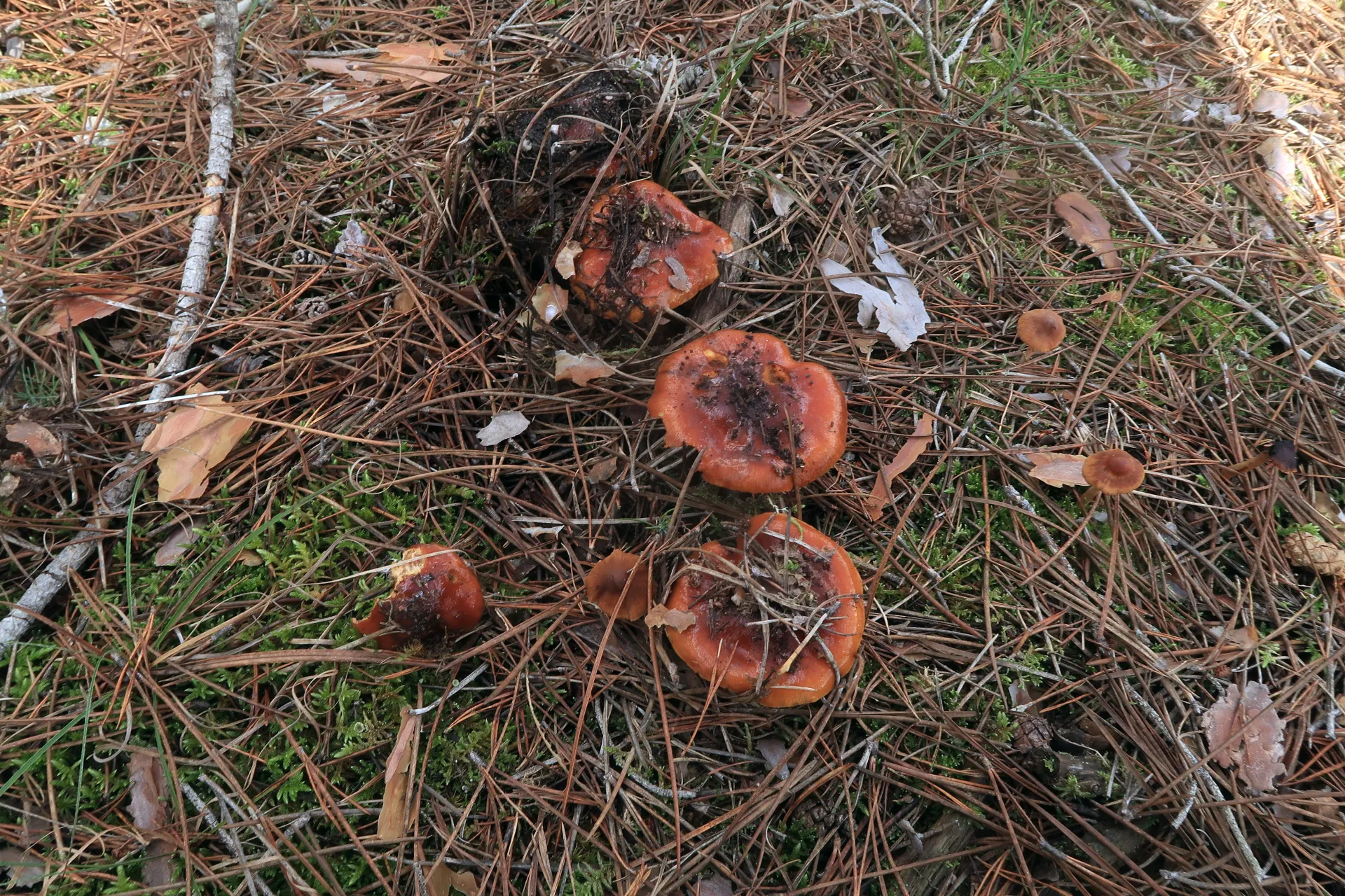
Cortinarius mucosus
(Bull.) J.Kickx f.
Study description
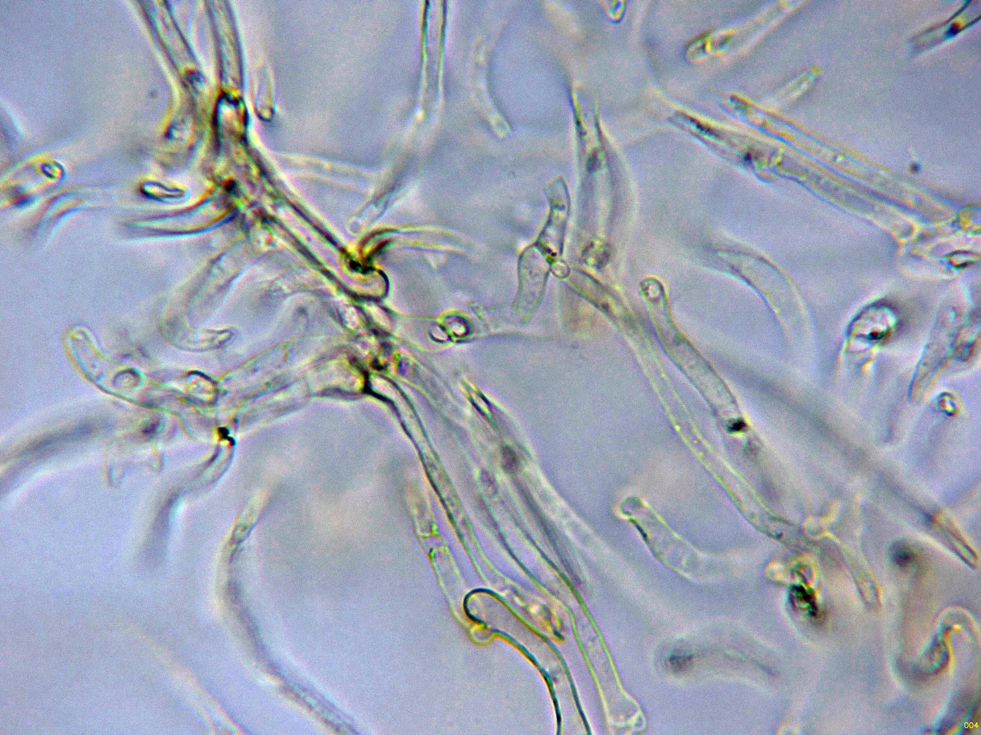
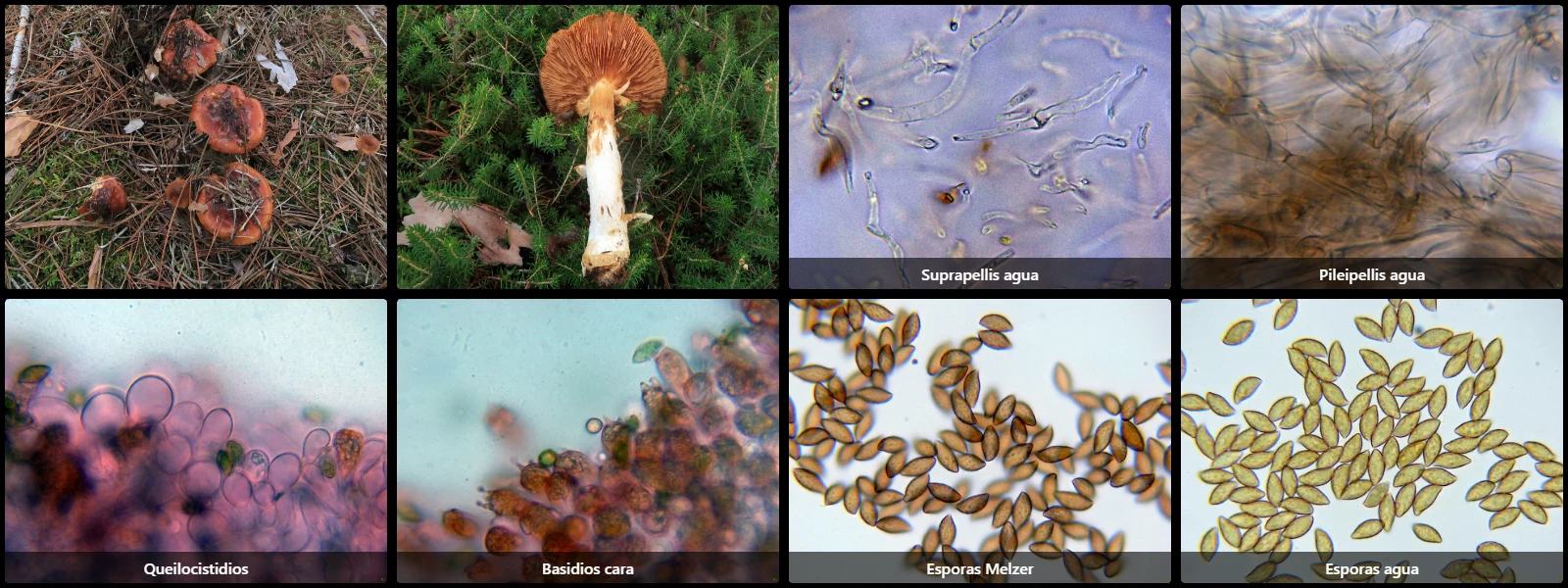
Filamentous suprapellis, formed by a layer of slender hyphae with ochraceous intracellular pigment, widely separated by gelatinization. Below this, broader hyphae arranged more or less parallel, similarly pigmented and clamped.
Lamellar edge completely covered by vesiculose to pyriform terminal cells acting as cheilocystidia, up to 40 µm wide.
Basidia cylindric-clavate, up to 33 µm long and 11 µm wide, with basal clamp connections, 4-spored.
Spores ellipsoidal to fusiform (almond-shaped), slightly verrucose-walled, yellow under the microscope with darker warts, a slightly eccentric apiculus, and moderately dextrinoid reaction in Melzer's reagent.
Herbarium material available: JTP2267/24.
Geographic references
Map generated from global occurrence data from GBIF. GBIF.
Study measurements
Values in Microns (µm)Macroscopic gallery
Microscopic gallery