


Candolleomyces candolleanus
(Fr.) D.Wächt. & A.Melzer
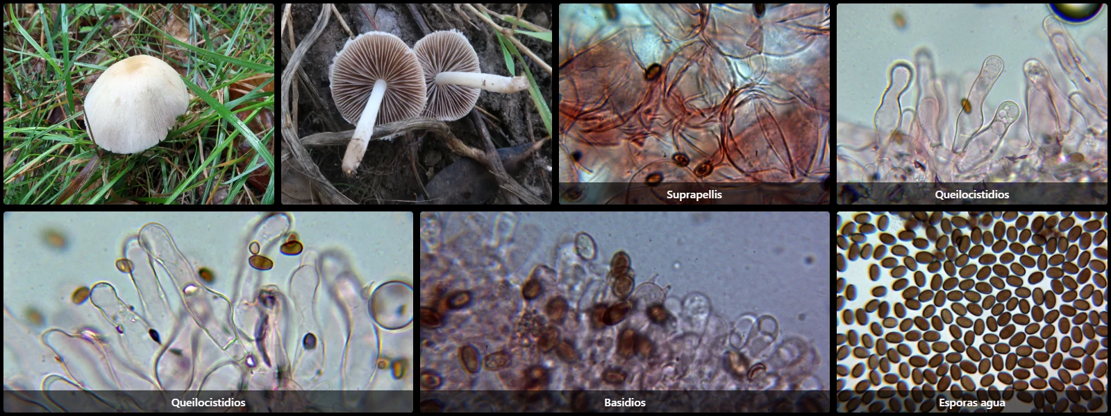
Study description
Suprapellis cellular (epithelioid), formed of spherical to slightly polygonal hyphae due to mutual compression, of considerable size and hyaline.
Cheilocystidia cylindrical to clavate, ventricose, hyaline, but containing lipid or oil droplets inside.
Pleurocystidia absent.
Basidia clavate and 4-spored.
Spores ellipsoidal, smooth, brown under the microscope, with a visible germ pore at one end.
Geographic references
Map generated from global occurrence data from GBIF. GBIF.
Study measurements
Values in Microns (µm)Macroscopic gallery
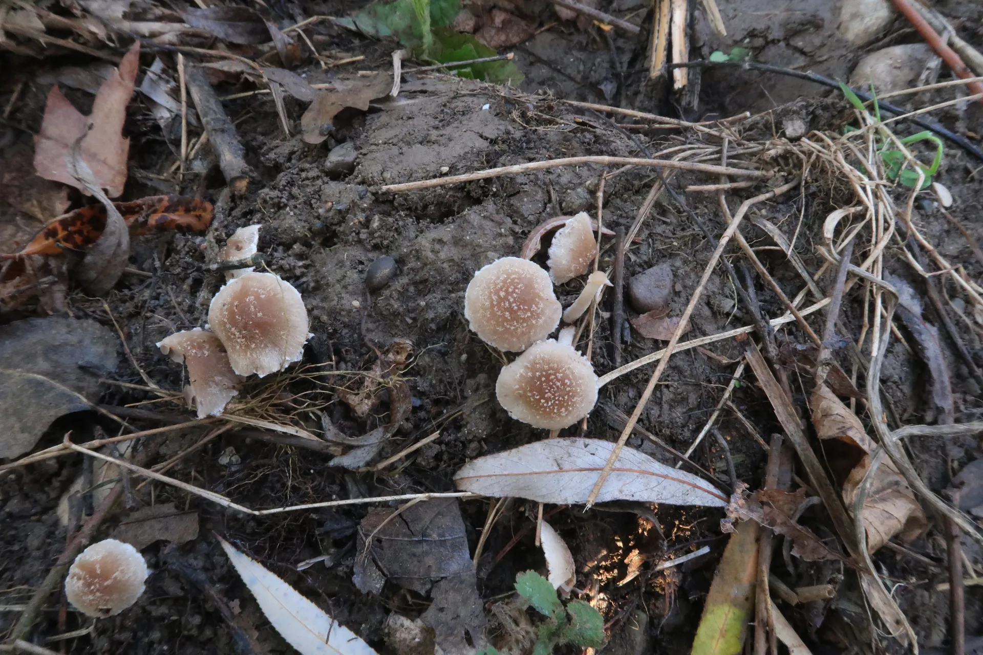
1 / 11Candolleomyces candolleanus - 1 - IMG_5845
Microscopic gallery
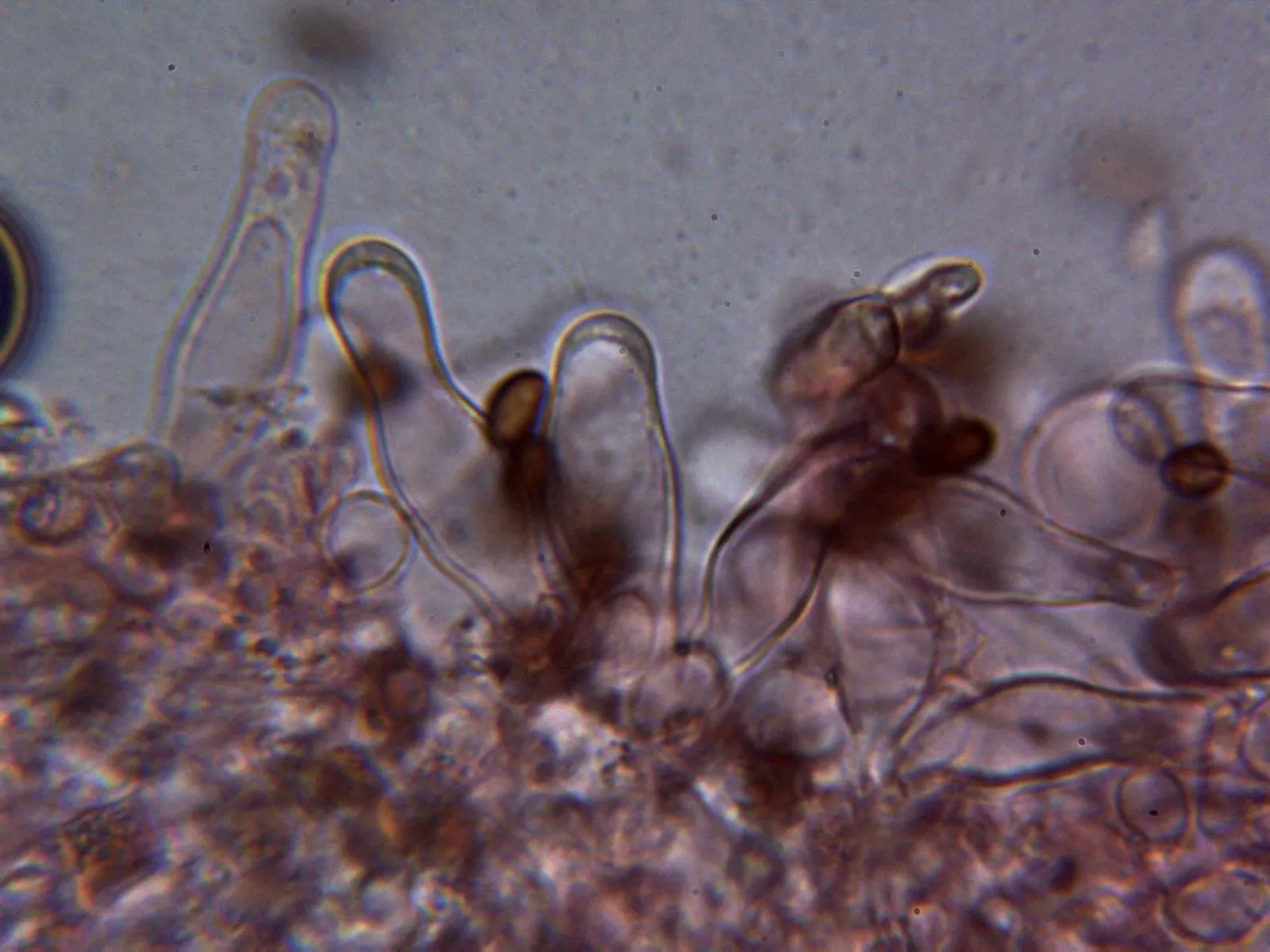
1 / 23Candolleomyces candolleanus - 10 - Queilocistidios 2