


Tympanis pini
Crous & R.K.Schumach.
Study description
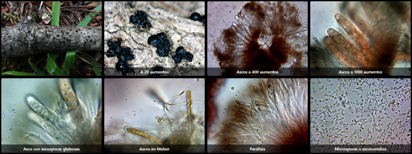
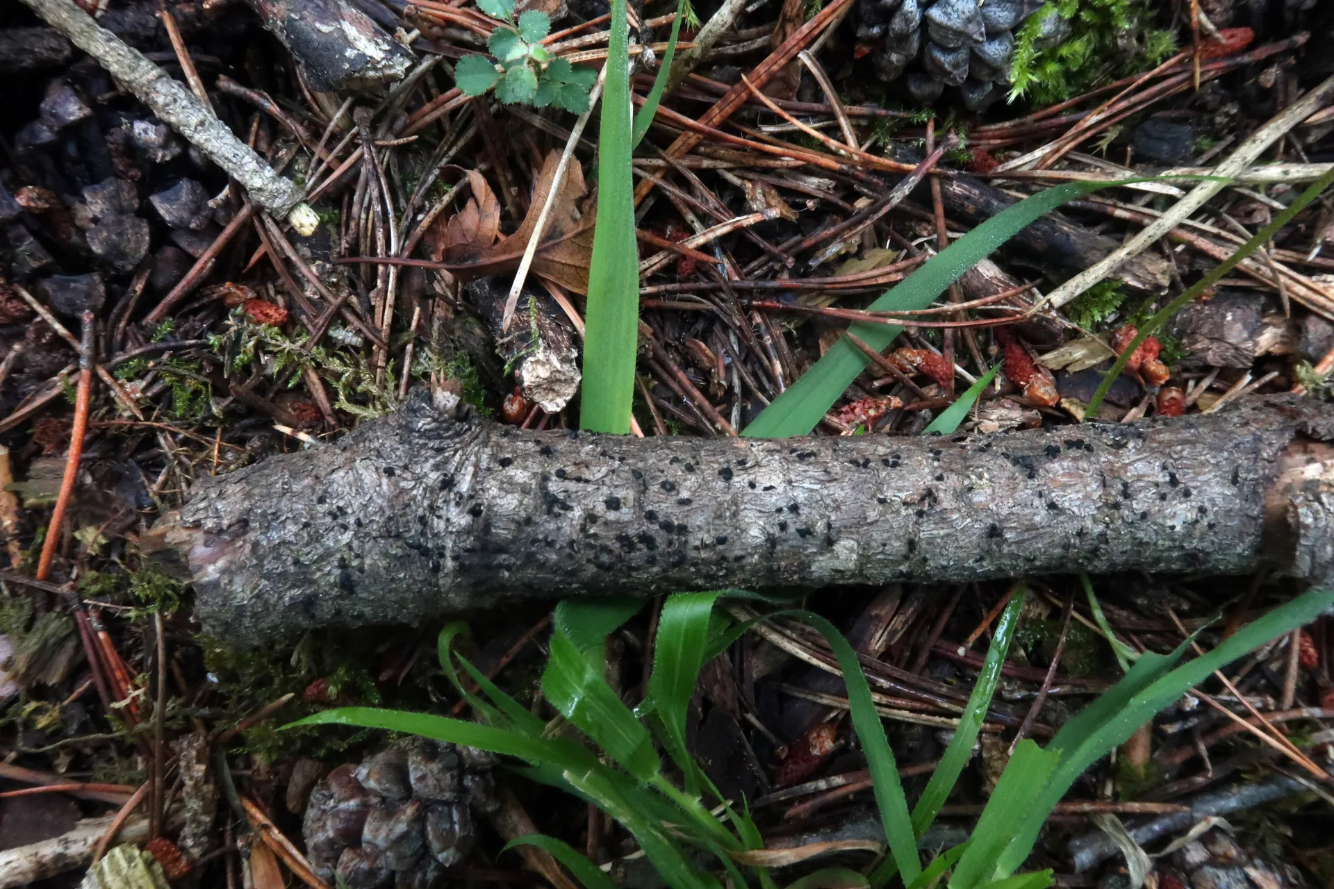
On a thin decaying branch on the ground of Pinus sylvestris, with erumpent ascomata in clustered grains, adopting a moriform to cerebriform appearance, black, and carbonaceous.
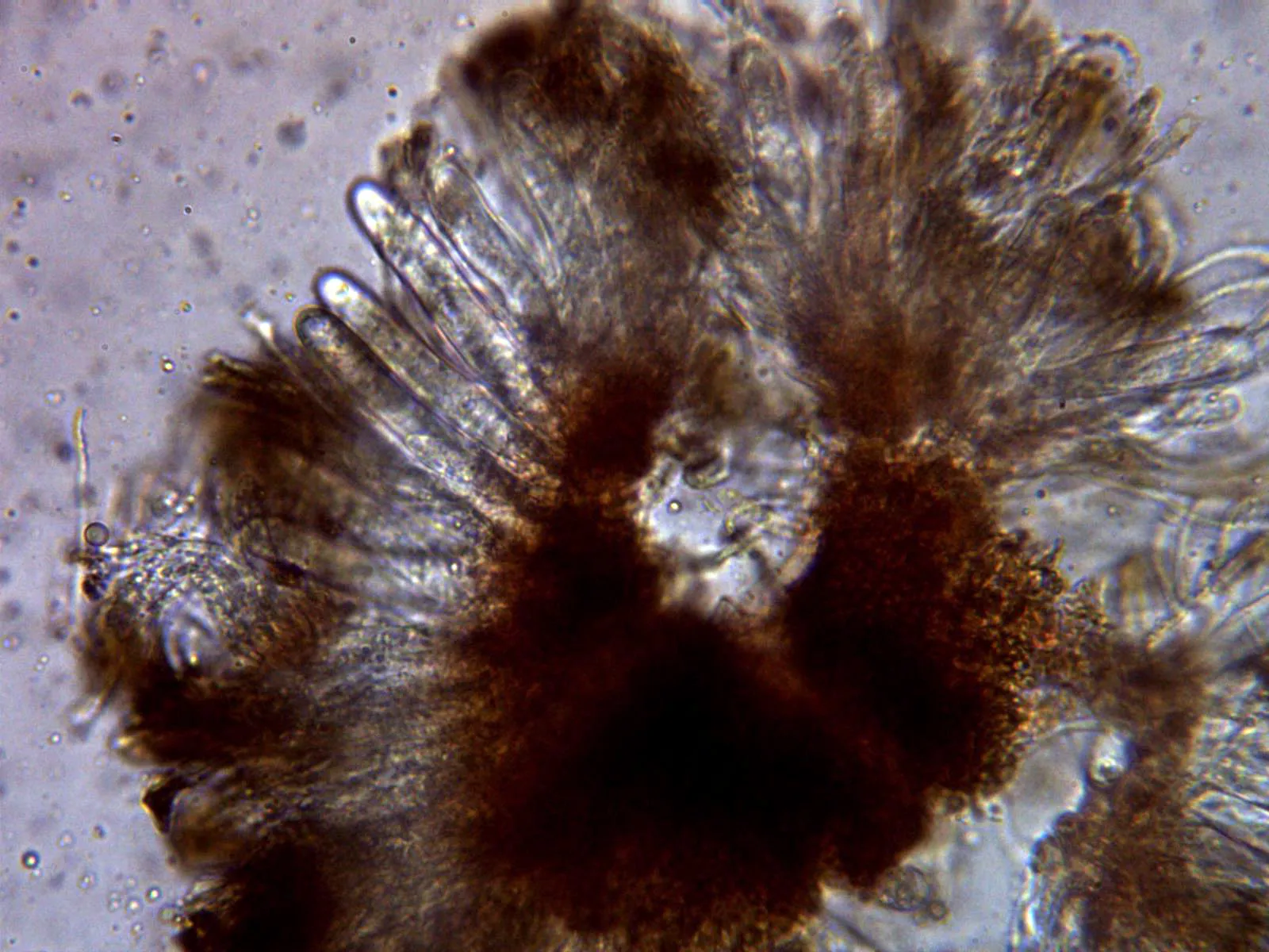
Asci cylindrical to clavate, bitunicate, 70 to 90 µm long, 9 to 15 µm wide, 8-spored in an initial stage, with globose ascospores that upon maturity act as mother cells, producing within themselves multiple microspores that the ascus releases externally, inamyloid in Melzer's reagent.
Paraphyses filiform, smooth, hyaline, multi-septate, 1.5 to 2 µm wide, embedded in a gelatinous matrix, projecting considerably above the level of the asci.
Microspores (or ascoconidia according to some authors), cylindrical, smooth, hyaline, thin-walled, 2.5 to 4 µm long, 0.8 to 1.3 µm wide, with homogeneous, very slightly granular contents.
Herbarium material available: JTP1925/21.
Geographic references
Map generated from global occurrence data from GBIF. GBIF.
Study measurements
Values in Microns (µm)Macroscopic gallery
Microscopic gallery
Genetic / Phylogenetic Study
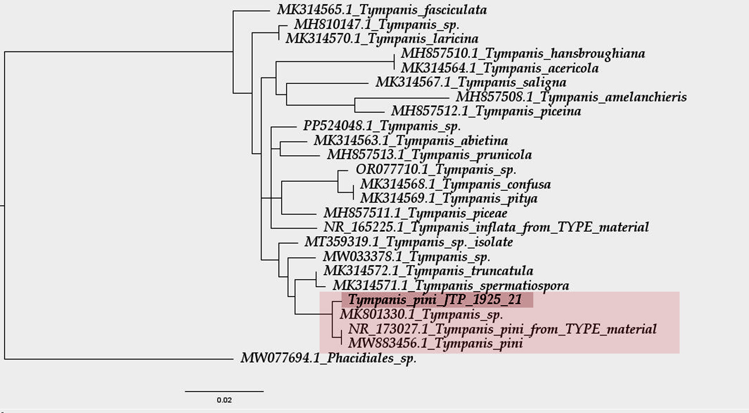
Phylogenetic Tree
Sequences and Data
Sequencing (Chromatogram)
Select a file to visualize the chromatogram