

Tubulicium vermiferum
(Bourdot) Oberw. ex Jülich
Study description
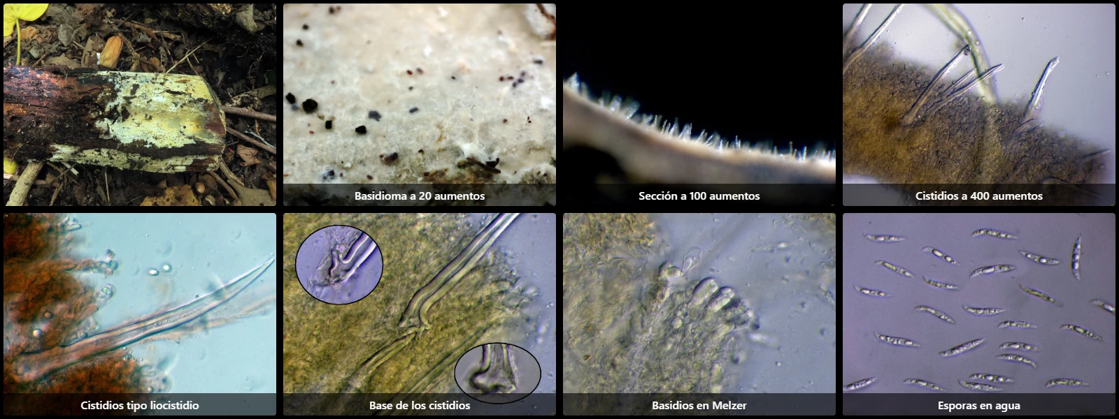
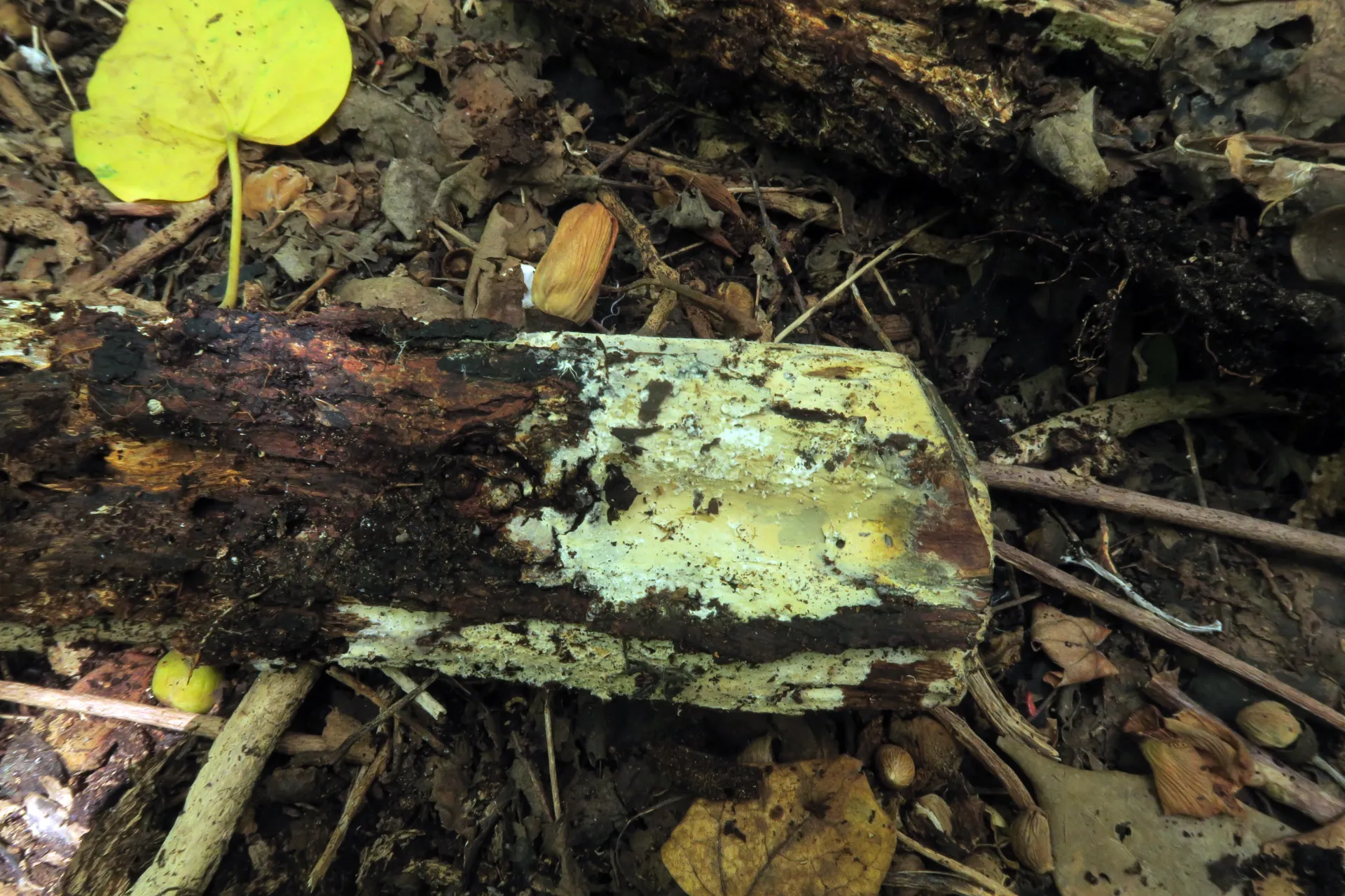
On decaying ground bark, with completely resupinate basidiomes, in irregular and separated patches, very thin, tightly adnate to the substrate, with a smooth and pubescent surface, whitish to pale yellowish, and with a diffuse, ill-defined growing margin.
Monomitic hyphal system, with very compacted subhymenial hyphae, difficult to dissociate, thin- to moderately thick-walled, 1.5 to 3 µm wide, septate with clamp connections at the septa.
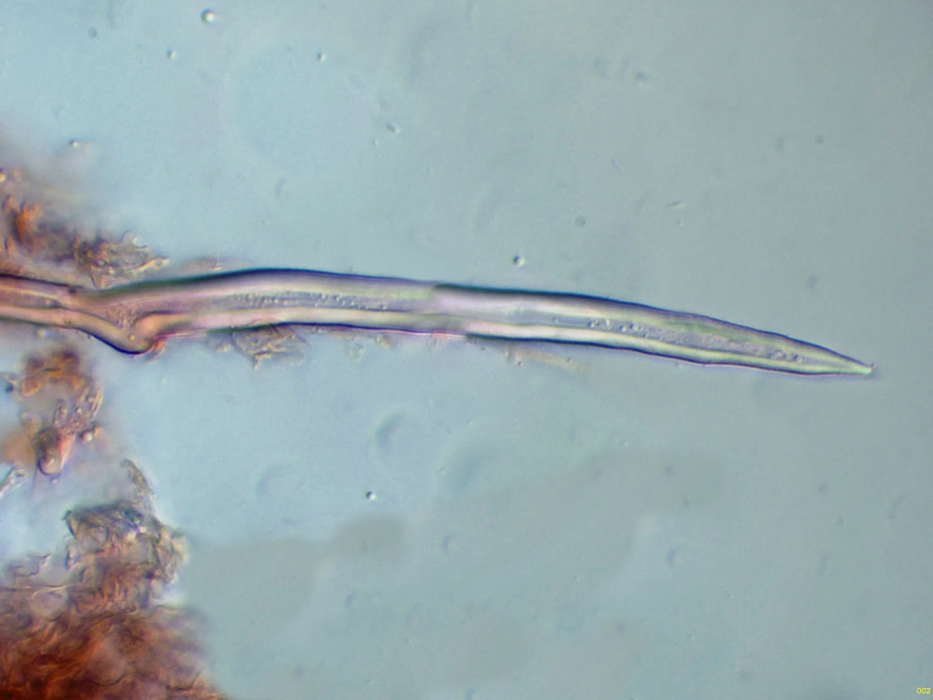
Cystidia very abundant, lyocystidium-type, conical, very thick-walled, with a narrow lumen, with a multi-rooted base (multifid), 35 to 125 µm long by 6 to 11 µm wide, and with basal enlargements of 8 to 20 µm.
Basidia subcylindrical to suburniform, 20 to 30 µm long by 7 to 10 µm wide, and tetrasporic, with sterigmata up to 8 µm.
Spores fusiform to sigmoid, smooth, hyaline, thin-walled, 18 to 25 µm long by 3.5 to 4.5 µm wide, with multiple oil drops of varying sizes inside, and inamyloid in Melzer's reagent.
Herbarium material available: JTP2170/24.
Geographic references
Map generated from global occurrence data from GBIF. GBIF.
Study measurements
Values in Microns (µm)Macroscopic gallery
Microscopic gallery