Tubeufia cerea
(Berk. & M.A.Curtis) Höhn.
Study description
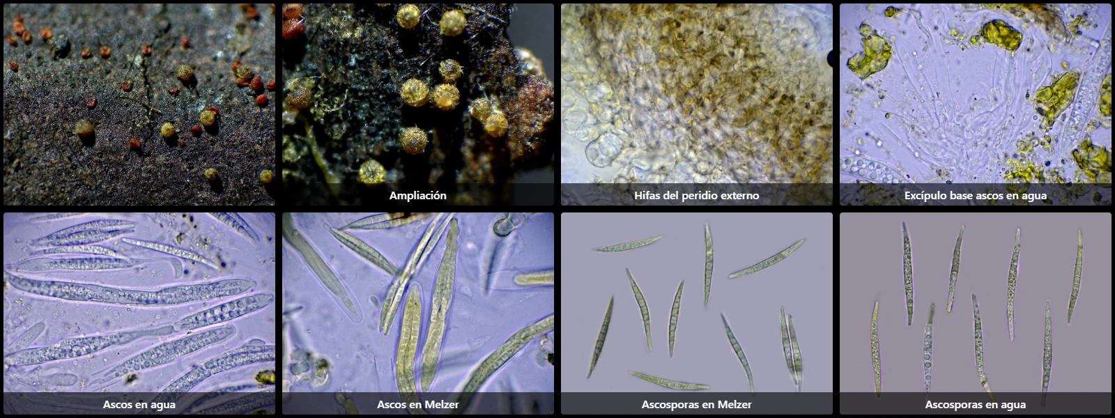
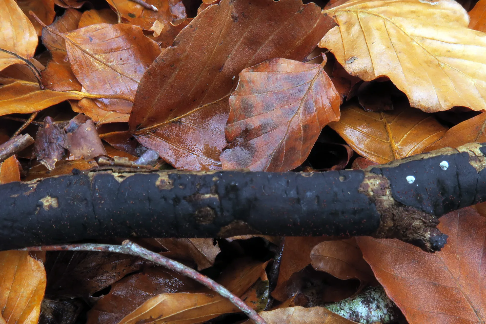
On decaying stromata of Diatrype, after remaining for an extended period in a moist chamber, with globose to pyriform ascomata, 0.2 to 0.3 mm in diameter, yellowish-brown, growing gregariously on the substrate.
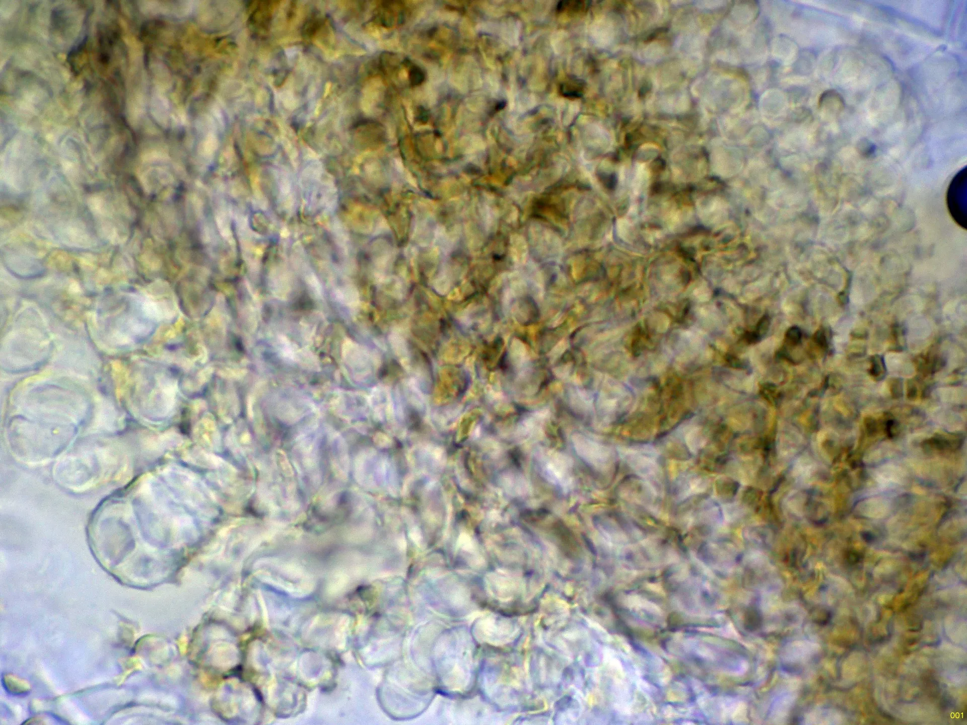
Ascoma wall hyphae of textura globulosa-angularis, thick-walled, densely compacted together (pseudoparenchymatous appearance), with abundant yellowish pigment inside.
Asci cylindrical, 8-spored, bitunicate, 70 to 100 µm long, 8 to 12 µm wide, spore arrangement within the ascus biseriate to fasciculate, and inamyloid in Melzer's reagent.
No paraphyses observed, although according to literature it may present in an early stage of development a hymenial tissue resembling pseudoparaphyses, filamentous, hyaline, 1 to 1.5 µm wide, branched, which tends to dissolve with the formation of the asci.
Ascospores filiform to fusiform, smooth, thin-walled, with 7 to 10 transverse septa, 45 to 60 µm long, 3.5 to 4.5 µm wide, containing very abundant oil droplets (guttules) inside.
Geographic references
Map generated from global occurrence data from GBIF. GBIF.
Study measurements
Values in Microns (µm)Macroscopic gallery
Microscopic gallery