

Tubaria romagnesiana
Arnolds
Study description
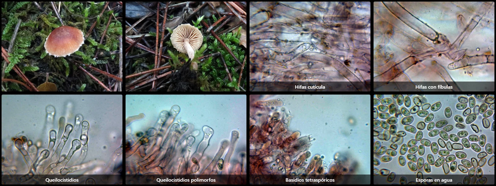
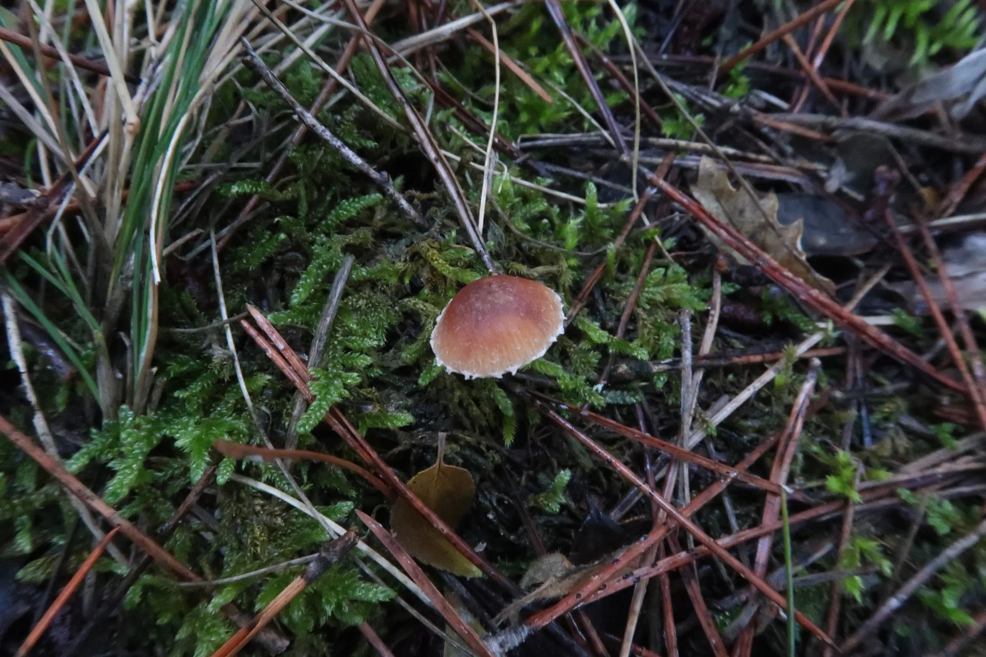
On decaying wood or soil plant debris, with a convex to flattened cap, 1 to 2.2 cm in diameter, with a smooth orange-brown surface darker in the center, whitish veil remnants at the margin, adnate whitish gills, a brown cylindrical stipe with whitish fibrils, a fungal odour, and a slightly bitter taste.
Filamentous cuticle formed by cylindrical hyphae arranged parallel to slightly intertwined with each other, 4 to 10 µm wide, with a light brown pigment inside, often encrusted on the surface, septate with clamp connections.
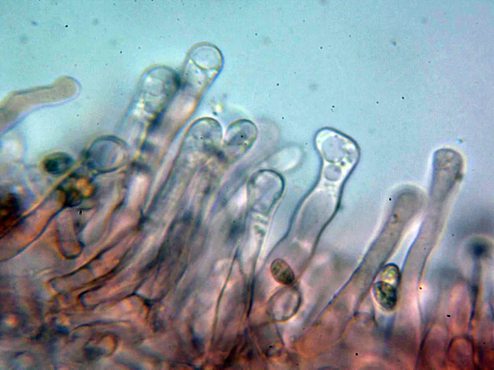
Cheilocystidia abundant, cylindrical to lageniform, sinuous, often capitate, slightly thick-walled, 45 to 90 µm long, with a maximum width of 6 to 15 µm.
Without pleurocystidia.
Lamellar trama parallel, with hyphae 7 to 15 µm wide.
Basidia clavate, 20 to 30 µm long by 6 to 8 µm wide, with basal clamp connections and tetrasporic.
Spores cylindric-ellipsoidal, smooth, thin- to moderately thick-walled, pale greyish-yellow under the microscope, with a barely noticeable eccentric apiculus at one end, 6.5 to 8 µm long by 4 to 5 µm wide, with oil drops (guttules) of varying sizes inside, and inamyloid in Melzer's reagent.
Geographic references
Map generated from global occurrence data from GBIF. GBIF.
Study measurements
Values in Microns (µm)Macroscopic gallery
Microscopic gallery