Tomentella punicea
(Alb. & Schwein.) J.Schröt.
Study description
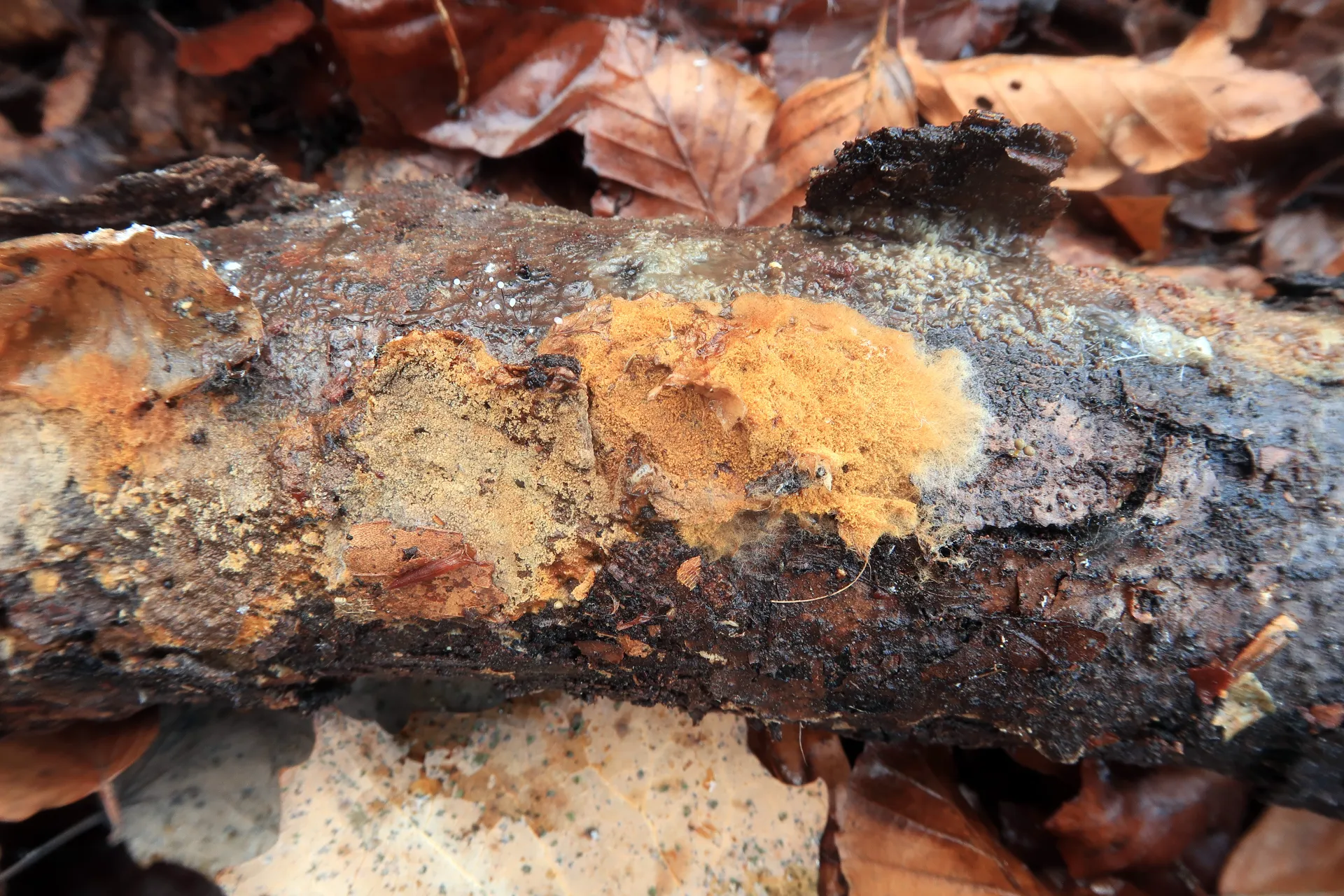
On decaying wood or bark on the ground, with completely resupinate basidiomes, granular ochraceous-brown surface, up to 1 mm thick, easily separable from the substrate, and a diffuse, whitish growing margin.
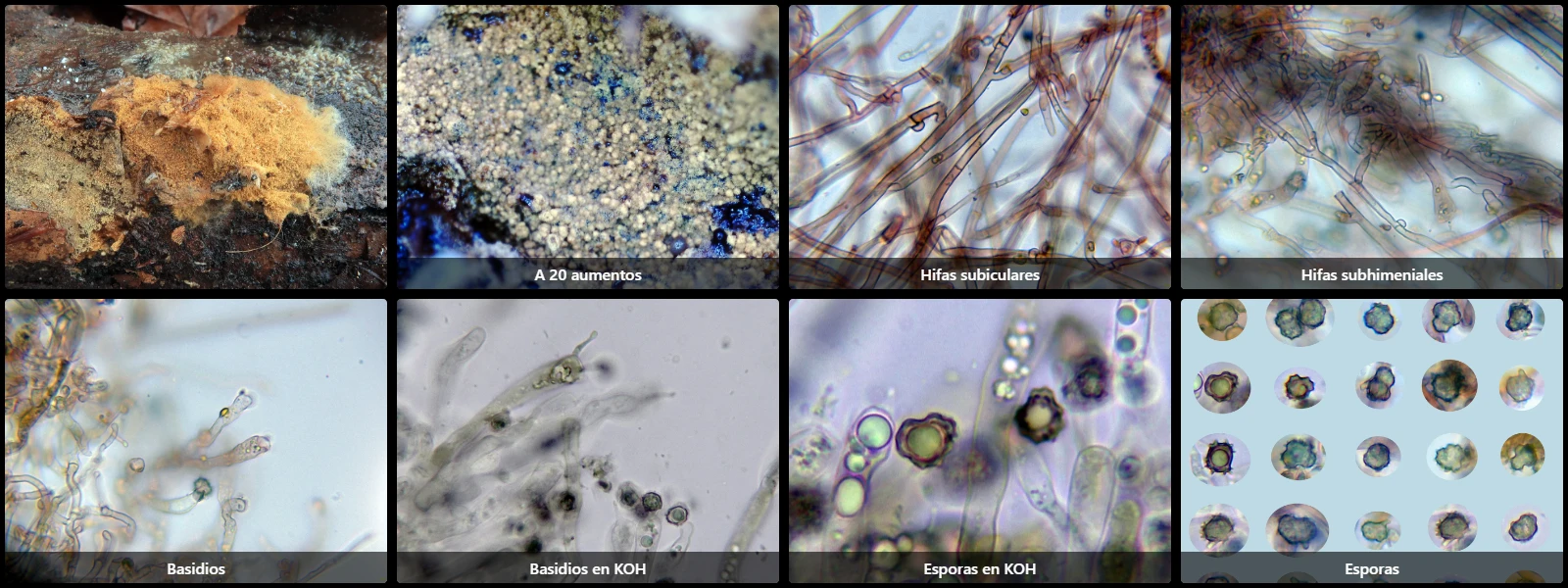
Subicular hyphae thin- to slightly thick-walled, 2 to 5 µm wide, pale yellow under the microscope in water, with some branching, septate, with clamp connections.
Subhymenial hyphae similar, hyaline, 3 to 4 µm wide, septate and clamped.
Without cystidia.
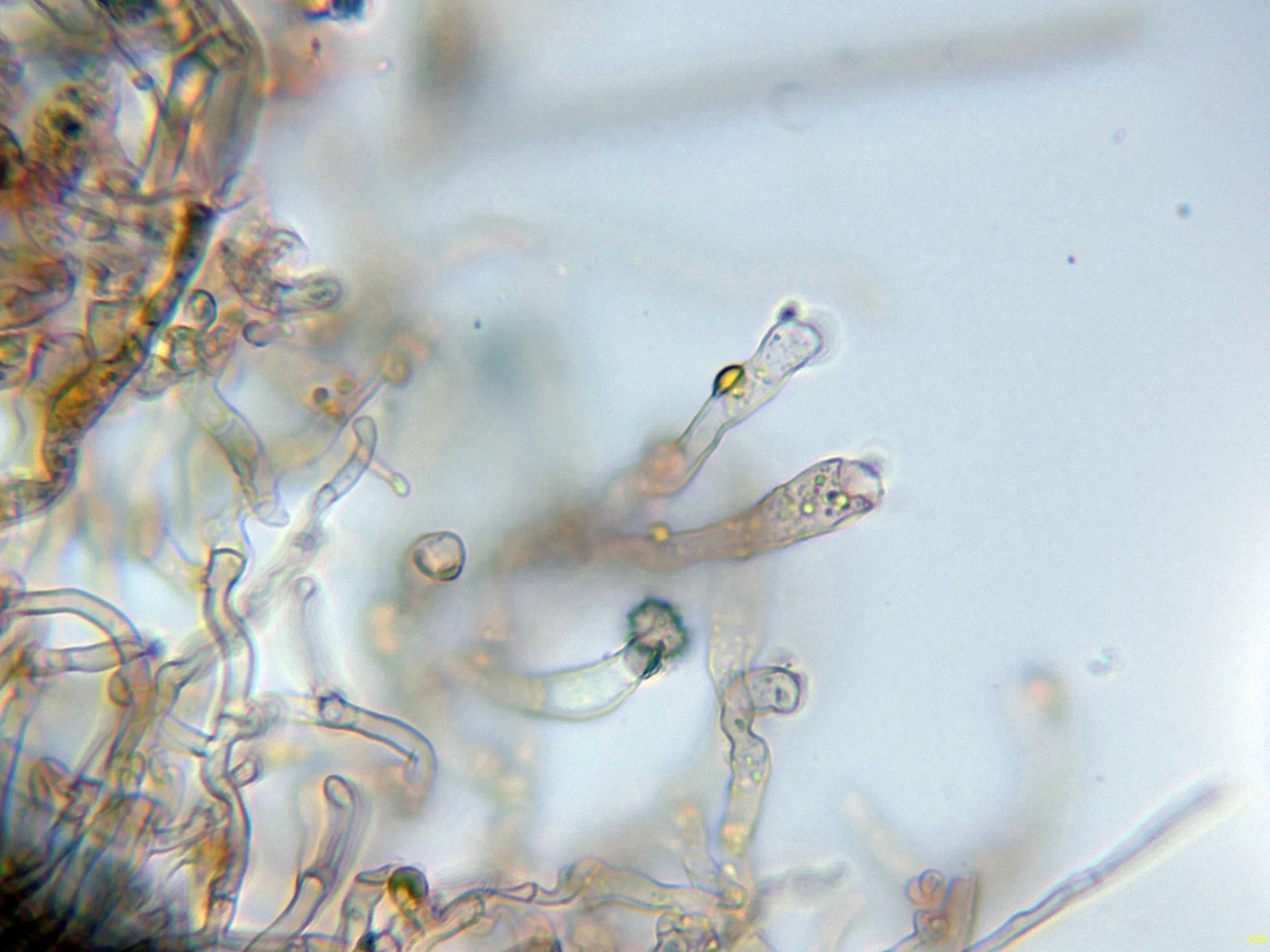
Basidia cylindric-clavate, sinuous, 40 to 60 µm long, 6 to 8 µm wide, often with yellowish granules inside, with basal clamp connections, 4-spored, with sterigmata up to 7 µm.
Spores very irregular in outline, lobed, echinulate, thick-walled, pale yellow under the microscope, 6 to 8.5 µm long excluding ornamentation, 5 to 8 µm wide, with spines 0.5 to 1 µm, containing oil droplets inside, inamyloid in Melzer's reagent.
Herbarium material available: JTP2313/24.
Geographic references
Map generated from global occurrence data from GBIF. GBIF.
Study measurements
Values in Microns (µm)Macroscopic gallery
Microscopic gallery