Tomentella asperula
(P.Karst.) Höhn. & Litsch.
Study description
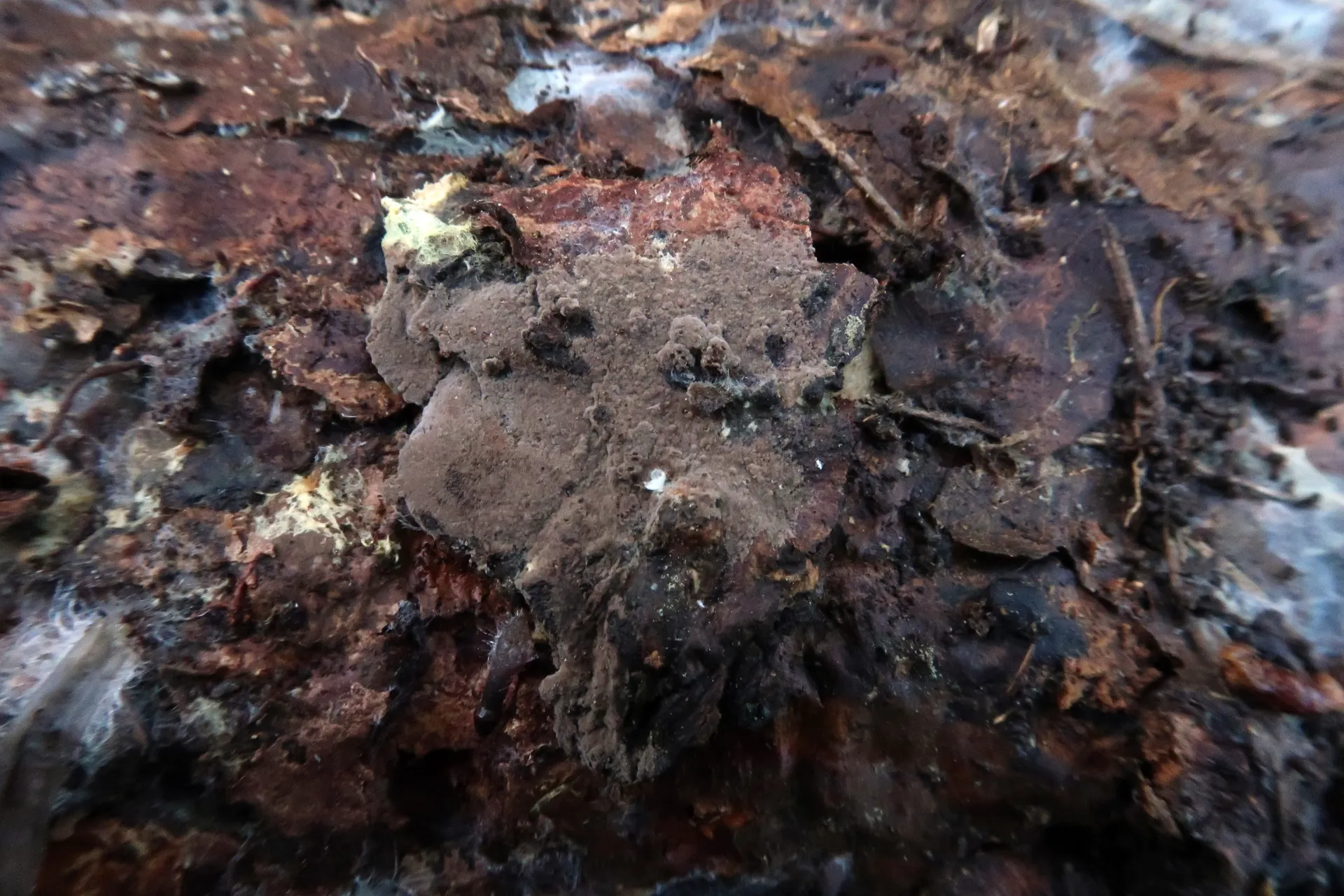
On decaying plant debris on the ground, with resupinate basidiomes, easily separable from the substrate, 0.5 to 1 mm thick, smooth to granulose surface when observed with a good hand lens, brown, with abundant rhizomorphs in the subiculum visible in the marginal zone.
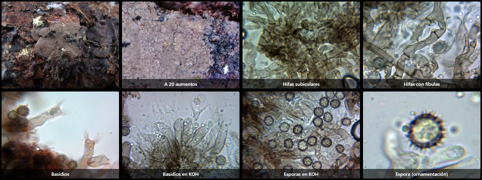
Monomitic hyphal system with moderately thick-walled subicular hyphae, 4 to 6 µm wide, containing brown pigment inside, branching at right angles, septate, with clamp connections.
Subhymenial hyphae thin- to slightly thick-walled, 3 to 5 µm wide, hyaline or with slight pale brown pigment inside, septate, with clamp connections.
Without cystidia.
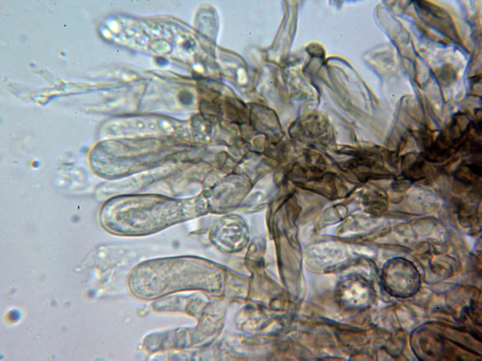
Basidia subcylindrical to clavate, sinuous, 40 to 75 µm long, 9 to 12 µm wide in the apical zone, sterigmata up to 9 µm long, with basal clamp connections, 4-spored.
Spores globose to subglobose, with regular outline, moderately thick-walled, yellowish-brown under the microscope, 8.5 to 10 µm long, 7.5 to 9.5 µm wide, ornamented with spines up to 2 µm high, with a faint apiculus, turning greenish in KOH, inamyloid in Melzer's reagent.
Geographic references
Map generated from global occurrence data from GBIF. GBIF.
Study measurements
Values in Microns (µm)Macroscopic gallery
Microscopic gallery