Thecotheus pelletieri
(P.Crouan & H.Crouan) Boud.
Study description
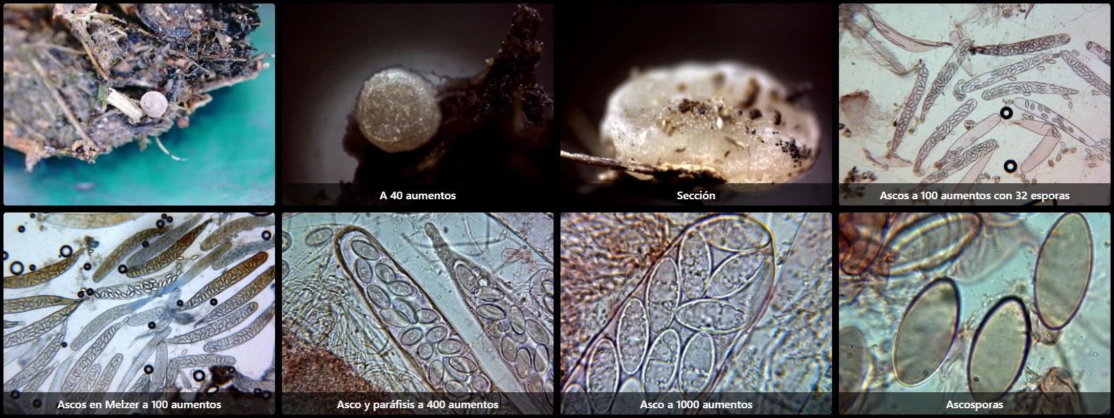
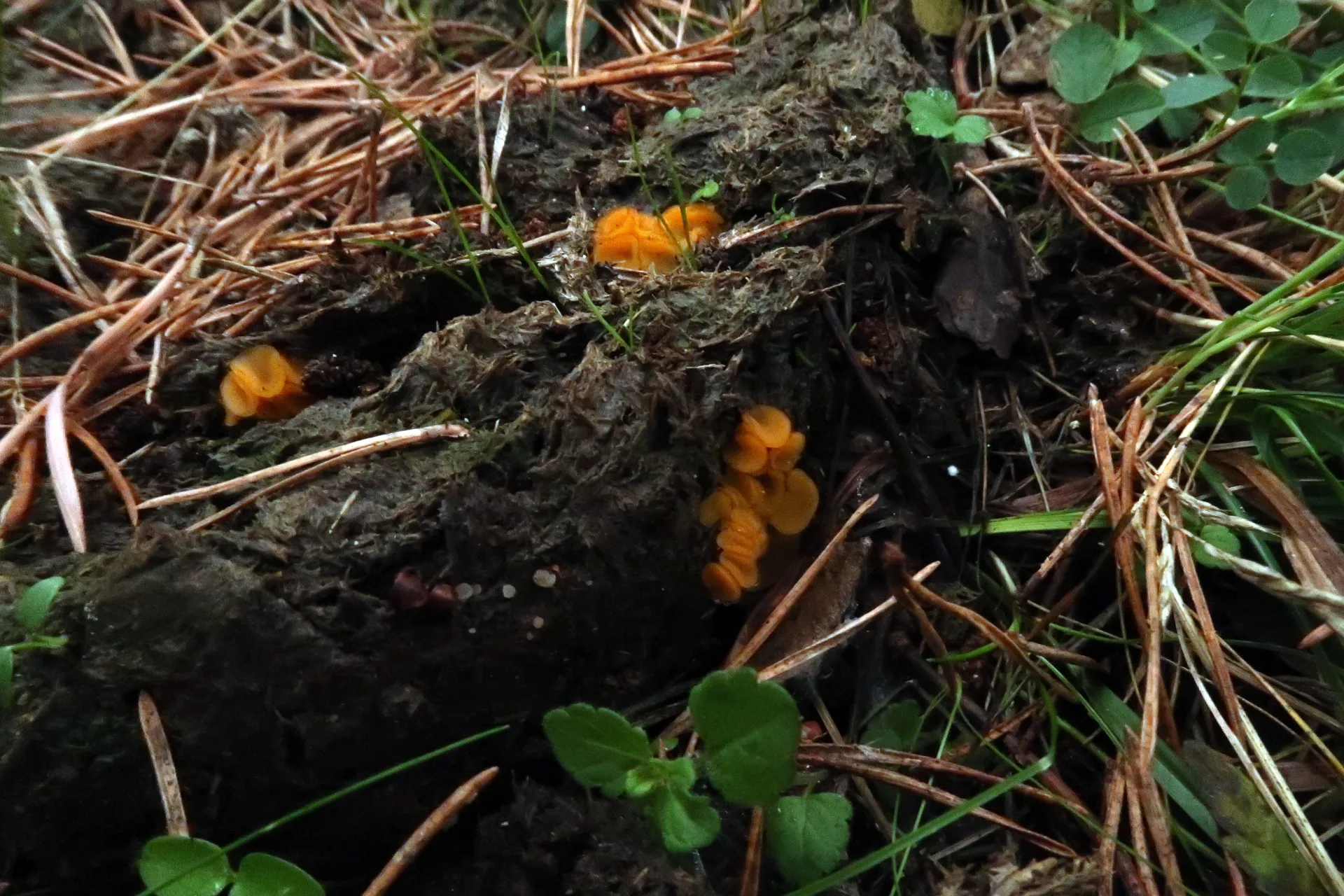
On decaying cow dung, with small subglobose to discoid apothecia, pale cream-brown, 0.5 to 0.9 mm in diameter and 0.5 mm high.
Asci readily visible even at 100× magnification, cylindrical-clavate, operculate, with an attenuated base, 382 to 418 µm long and 46 to 54 µm wide, with amyloid walls in Melzer's reagent, and mostly containing 32 spores inside.
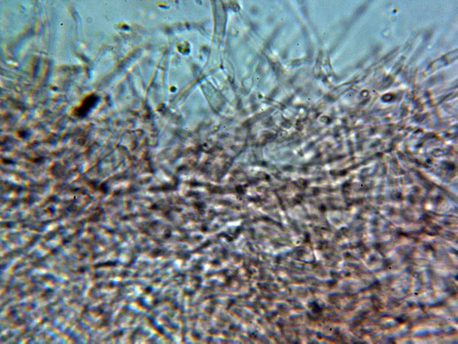
Interascal tissue functioning as paraphyses, filamentous, septate, hyaline, branched, immersed in a gelatinous matrix, 2 to 3 µm wide, and projecting well below the level of the asci.
Ascospores ellipsoidal to subfusiform, with slightly attenuated ends, thick-walled (the wall being much more distinct in Melzer's), 30 to 35 µm long, 14 to 18 µm wide, with granular to smooth contents depending on the stage of development, and inamyloid in Melzer's reagent.
Geographic references
Map generated from global occurrence data from GBIF. GBIF.
Study measurements
Values in Microns (µm)Macroscopic gallery
Microscopic gallery