Splanchnonema platani
M.E.Barr
Study description
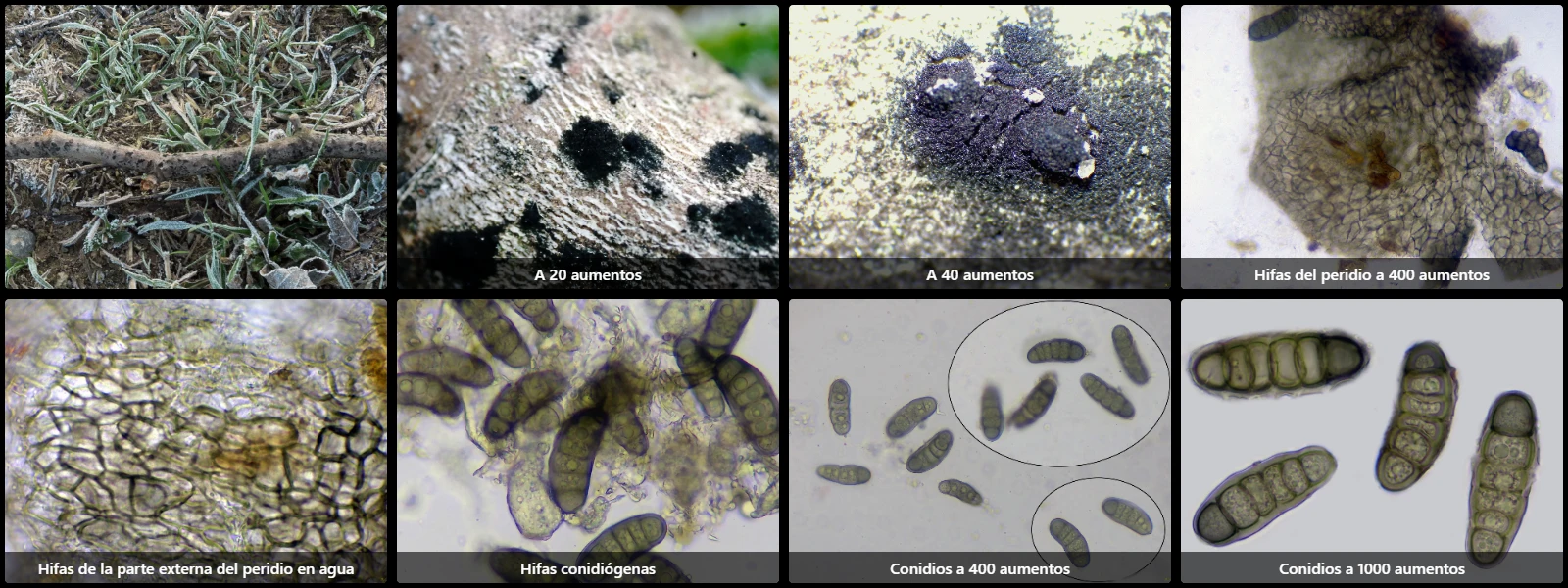
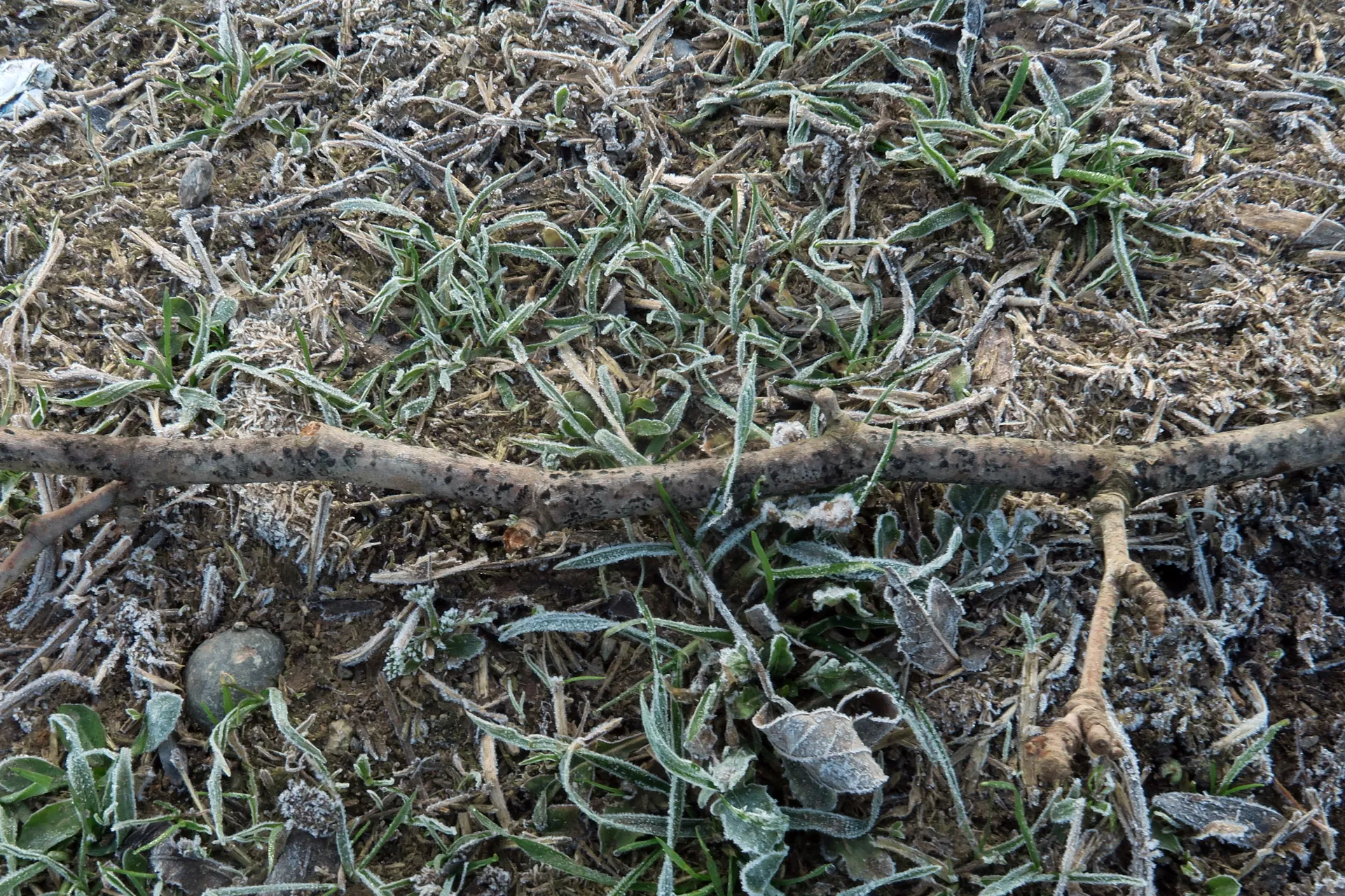
On a slender fallen twig of Platanus hispanica on the ground, with irregularly shaped erumpent grain-like pycnidia deeply immersed in tissue, 0.35 to 0.6 mm across, black, staining the surrounding area black upon discharging conidia.
This study corresponds to the anamorph (asexual or conidial stage) of the species.
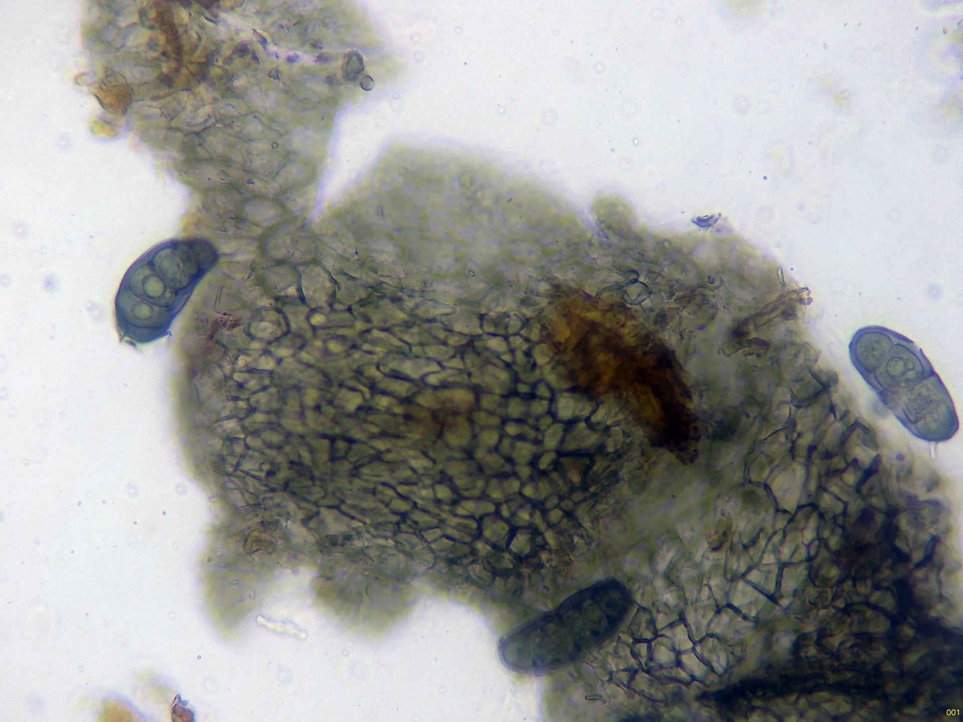
Peridial hyphae with an outer layer of thick-walled hyphae of textura angularis, 10 to 30 µm in diameter, with brownish intracellular pigment, and an inner layer of thin-walled hyphae of similar morphology but completely hyaline.
Conidiogenous cells on the inner perithecial wall, barely visible, developing from the innermost layer, cylindrical to subcylindrical and completely hyaline.
Conidia cylindrical-ellipsoidal to slightly allantoid, smooth- and thick-walled, brown with greenish tints under the microscope, 45 to 60 µm long, 15 to 22 µm wide, with 5 thick transverse septa, generally enveloped in a gelatinous sheath and containing oil droplets (guttules) inside.
Herbarium material available: JTP2719/26.
Geographic references
Map generated from global occurrence data from GBIF. GBIF.
Study measurements
Values in Microns (µm)Macroscopic gallery
Microscopic gallery