Seiridium marginatum
Nees
Study description
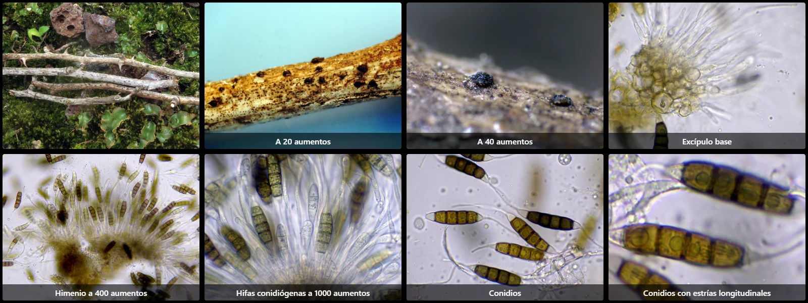
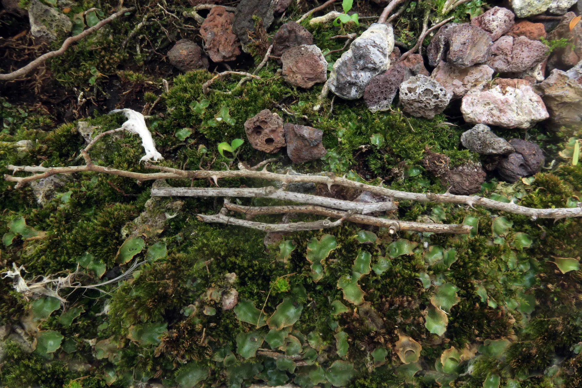
On slender twigs still attached to a Crataegus sp. shrub, with fruit bodies appearing as small erumpent black pustules that rupture the bark, sprouting scattered across the surface.
This study corresponds to the asexual or conidial stage of the species.
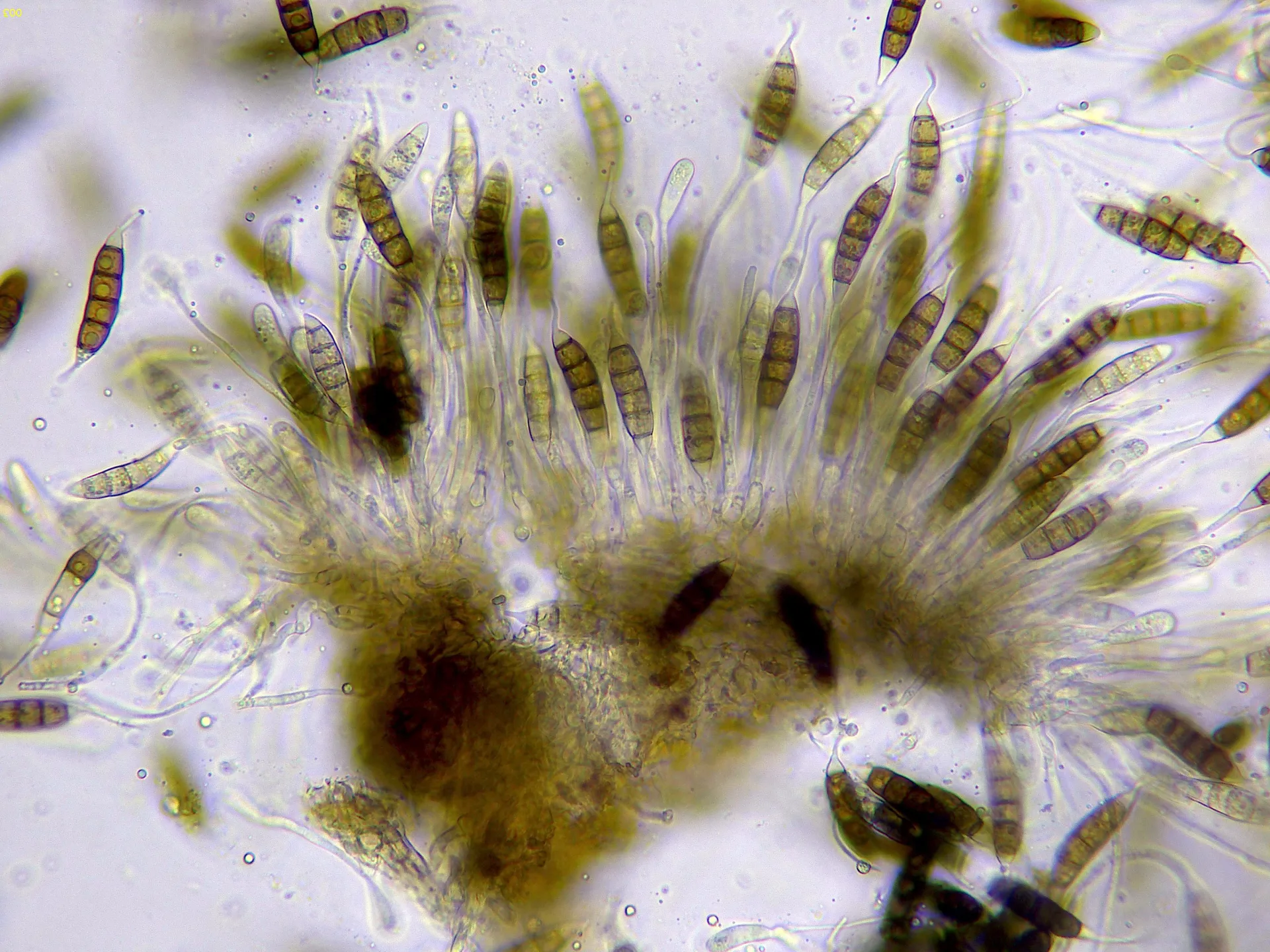
Inside the acervuli, conidiogenous cells are arranged more or less radially; these conidiogenous cells are hyaline, simple or with a few branches, cylindrical, slightly tapered at the apex where they generate conidia singly.
Conidia fusiform, with five transverse septa dividing the conidium into 6 cells (terminal cells hyaline, the 4 median cells greenish brown in water under the microscope), 38 to 47 µm long, 9 to 11 µm wide, with hyaline appendages 40 to 50 µm long, and characteristic longitudinal striae running along their entire surface.
Herbarium material available: JTP2500/25.
Geographic references
Map generated from global occurrence data from GBIF. GBIF.
Study measurements
Values in Microns (µm)Macroscopic gallery
Microscopic gallery