Puccinia teucrii
Biv.
Study description
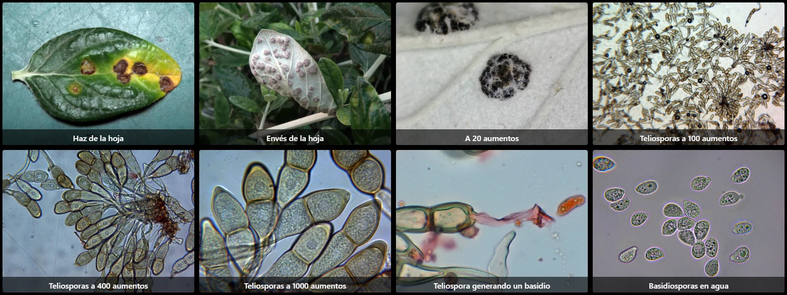
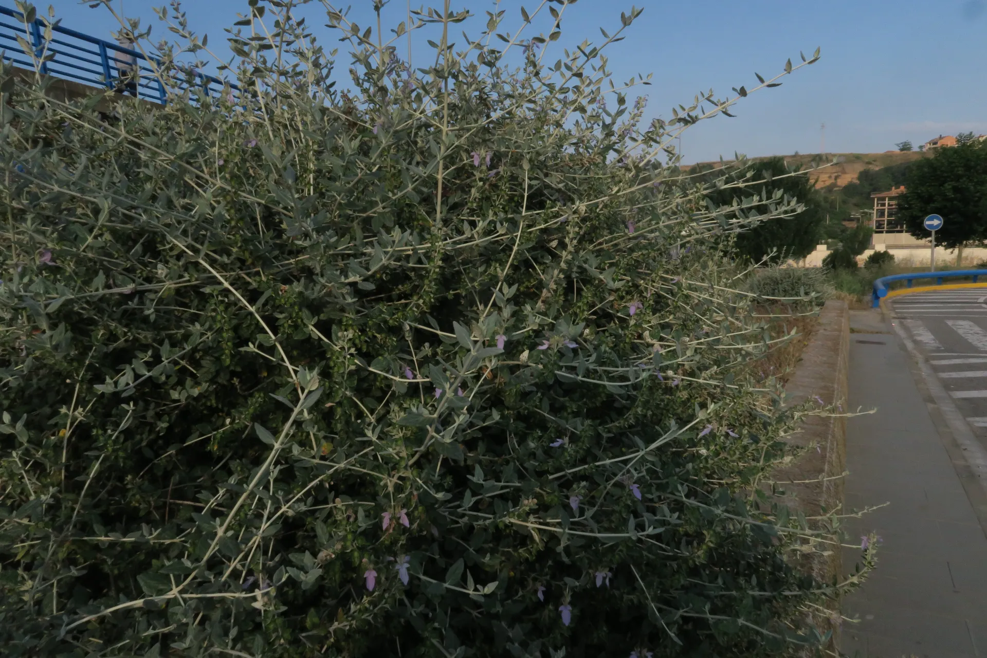
On living leaves of Teucrium fruticans, causing reddish-brown to yellowish necrotic spots on the upper surface and blackish pustules on the underside that rupture the leaf epidermis to release spores.
This study corresponds to the telial stage of the species, based on two collections: one in my hometown (Balaguer) and another in a public park in Barcelona.
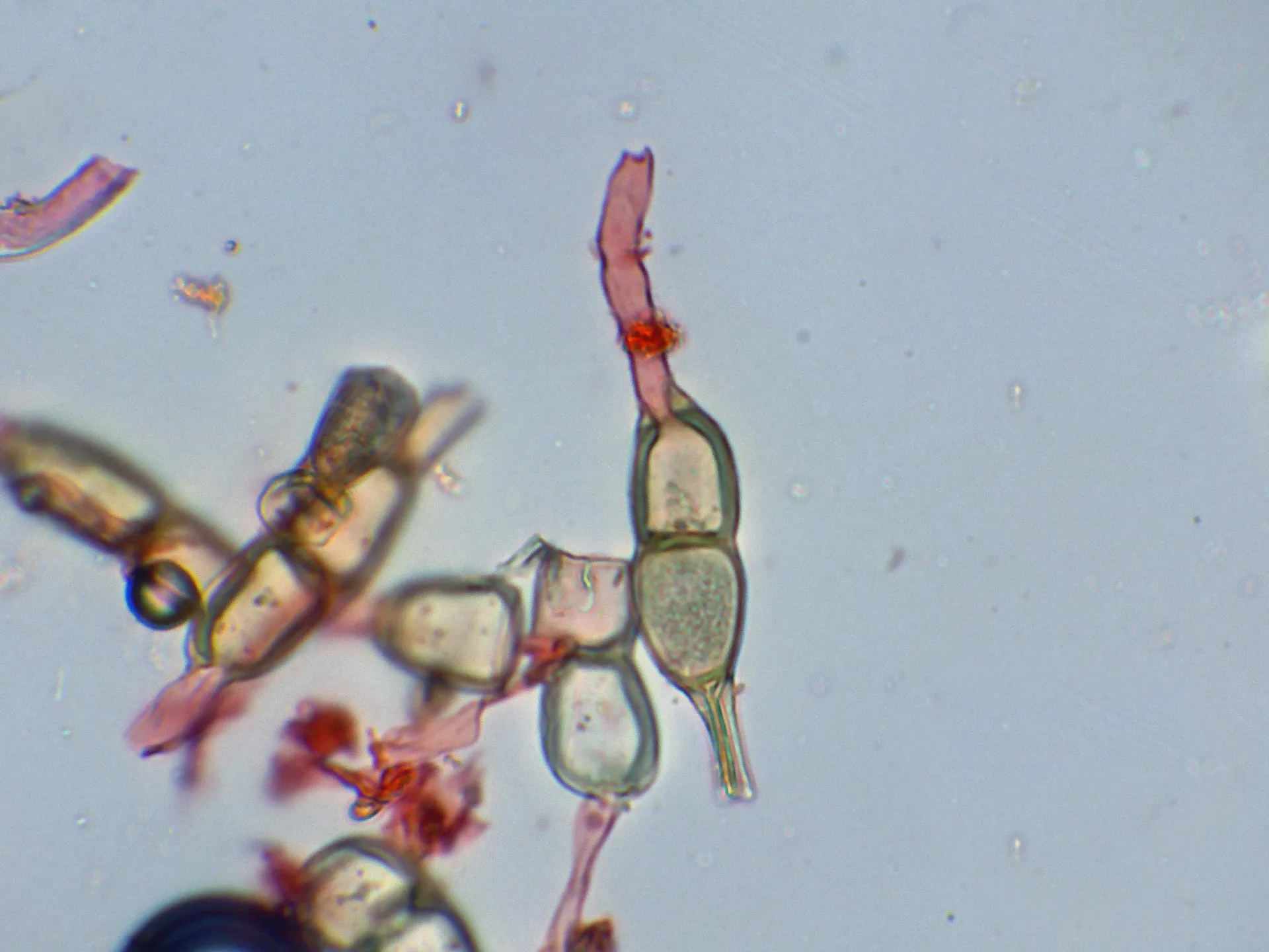
Teliospores ellipsoidal, slightly fusiform to clavate, smooth-walled, with a thick central septum and marked constriction at the septum, brown in the upper cell to more hyaline in the lower cell, 35 to 55 µm long, 15 to 20 µm wide, wall thickness 2 to 3 µm, with an apical thickening up to 5 µm, and a long hyaline pedicel 80 to 140 µm long.
Basidiospores ellipsoidal to amygdaliform, smooth, hyaline, thin-walled, 12 to 17.5 µm long, 8 to 12 µm wide, with a pointed end corresponding to the apiculus, and containing minute oil droplets (guttules) inside.
Geographic references
Map generated from global occurrence data from GBIF. GBIF.
Study measurements
Values in Microns (µm)Macroscopic gallery
Microscopic gallery