Psathyrella tephrophylla
(Romagn.) Bon
Study description
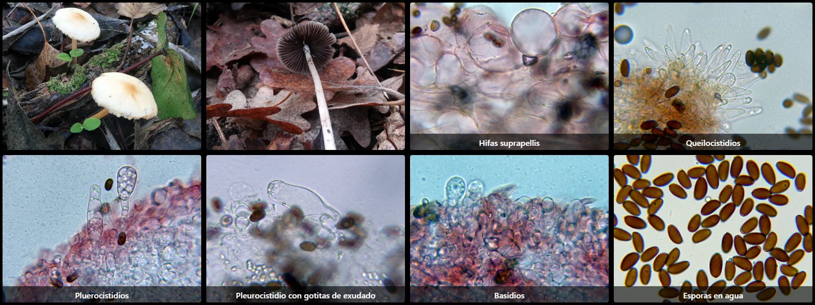
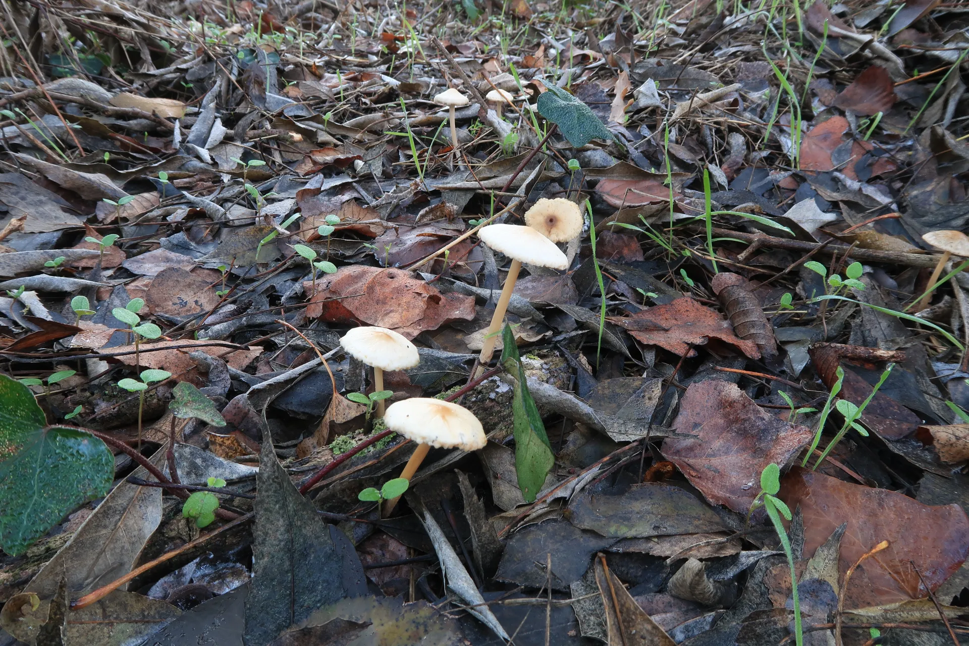
In forest clearings and along path edges on decaying buried woodchips or woody debris, with a conical-campanulate to flattened pileus 2 to 5 cm across, pale ochre with a darker disc, adnate violaceous-grey lamellae, and a cylindrical, hollow, fragile cream to whitish stipe.
Pileipellis cellular, composed of a layer of hyaline vesiculose to pyriform cells 30 to 55 µm long and 25 to 40 µm wide, clampless, overlying parallel cylindrical hyphae 8 to 15 µm wide.
Cheilocystidia fusiform to utriform, 30 to 45 µm long and 10 to 15 µm wide, intermixed with shorter pyriform cells, some bearing apical exudates of small hyaline droplets.
Pleurocystidia utriform to lageniform, 50 to 75 µm long and 12 to 17 µm wide.
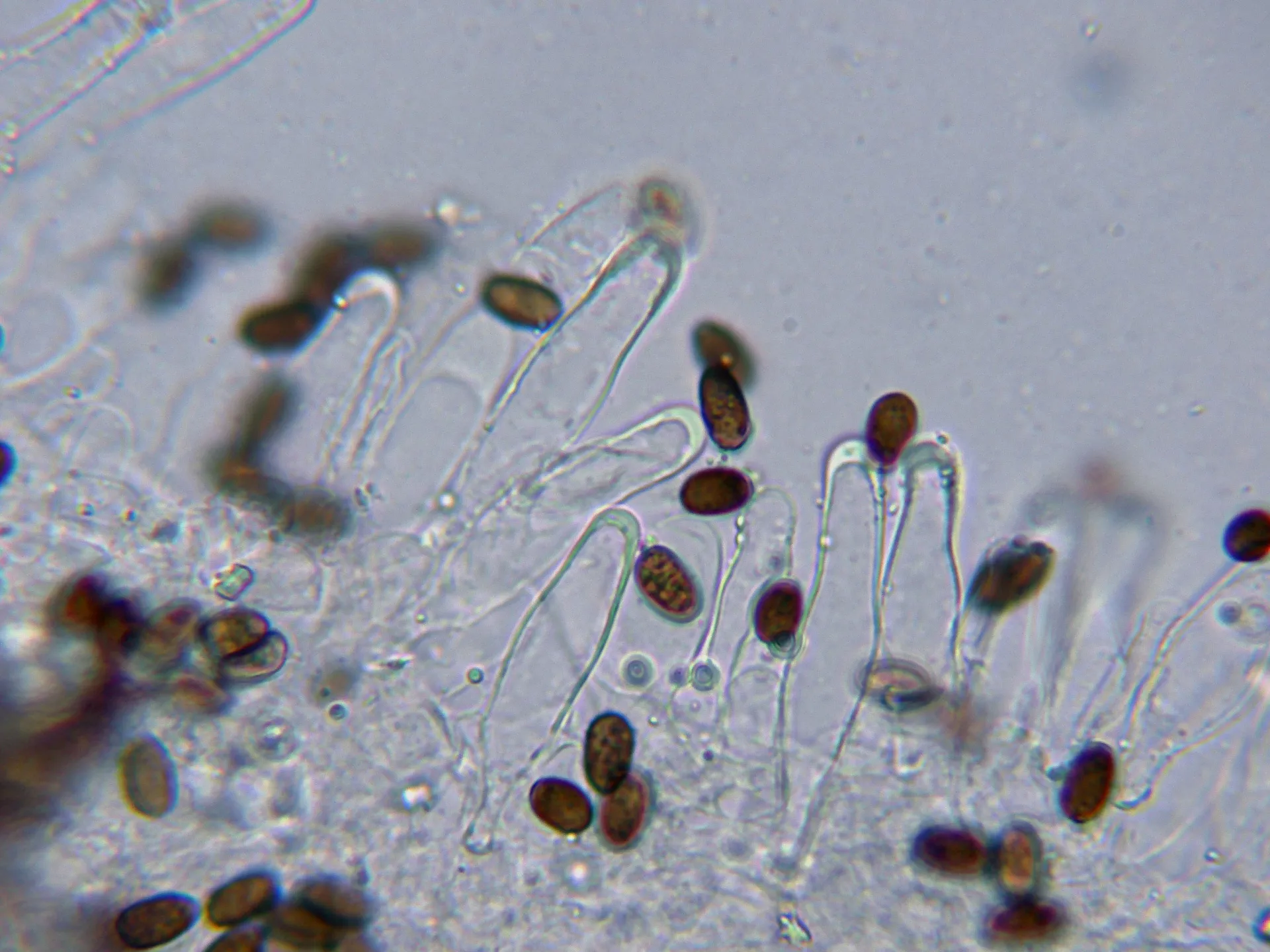
Basidia clavate, 15 to 20 µm long, 8 to 10 µm wide, clampless at the base, 4-spored.
Spores ellipsoidal, smooth, dark reddish-brown under the microscope, 9 to 12 µm long, 4.5 to 6.5 µm wide, with an inconspicuous eccentric apiculus at one end and a prominent central germ pore at the opposite pole.
Geographic references
Map generated from global occurrence data from GBIF. GBIF.
Study measurements
Values in Microns (µm)Macroscopic gallery
Microscopic gallery