Psathyrella pseudocorrugis
(Romagn.) Bon
Study description
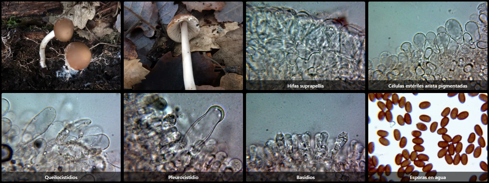
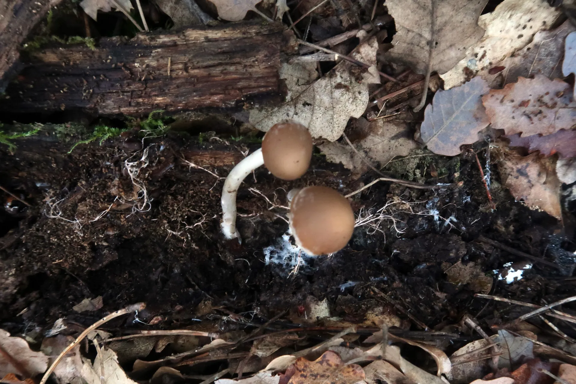
Under broadleaved trees on leaf litter or decaying fallen wood, with a hemispherical pileus expanding to convex or flattened, 2 to 6 cm across, brown to reddish-brown with whitish veil remnants along the margin, crowded lamellae with a pruinose edge, and a cylindrical whitish stipe with a pruinose surface.
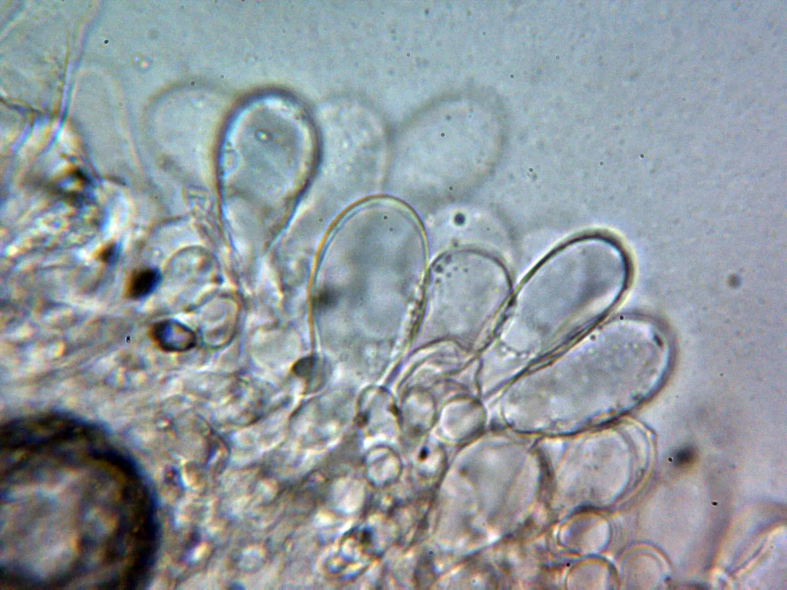
Pileipellis with a superficial layer of subglobose to pyriform cells 25 to 40 µm in diameter, hyaline or containing pale brownish intracellular pigment.
Cheilocystidia utriform to lageniform, mixed with numerous pyriform cells, with faint pigment inside, 25 to 50 µm long and 11 to 19 µm wide.
Pleurocystidia predominantly utriform to subcylindrical, some lageniform, 35 to 70 µm long and 11 to 20 µm wide.
Basidia clavate, 25 to 35 µm long, 7 to 10 µm wide, simple-septate at the base, 4-spored.
Spores ellipsoidal, smooth, medium brown under the microscope, 6.5 to 8 µm long, 4 to 5 µm wide, with an almost imperceptible apiculus at one pole and an evident germ pore at the opposite pole.
Geographic references
Map generated from global occurrence data from GBIF. GBIF.
Study measurements
Values in Microns (µm)Macroscopic gallery
Microscopic gallery