


Psathyrella microrhiza
(Lasch) Konrad & Maubl.
Study description
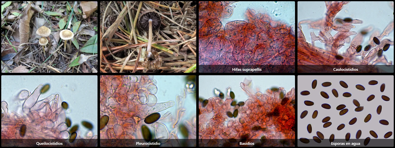
In soil among plant debris or decaying woody fragments, with a convex to flattened pileus 1.5 to 4 cm in diameter, pale brown to beige-brown, adnate to slightly decurrent lamellae, purplish-brown from sporulation, a cylindrical, hollow, fragile stipe, pale brown and whitish at the base, without distinct odour, and slightly herbaceous taste.
Pileipellis cellular, composed of hyaline globose, subglobose to pyriform cells 25 to 60 µm long and 15 to 40 µm wide.
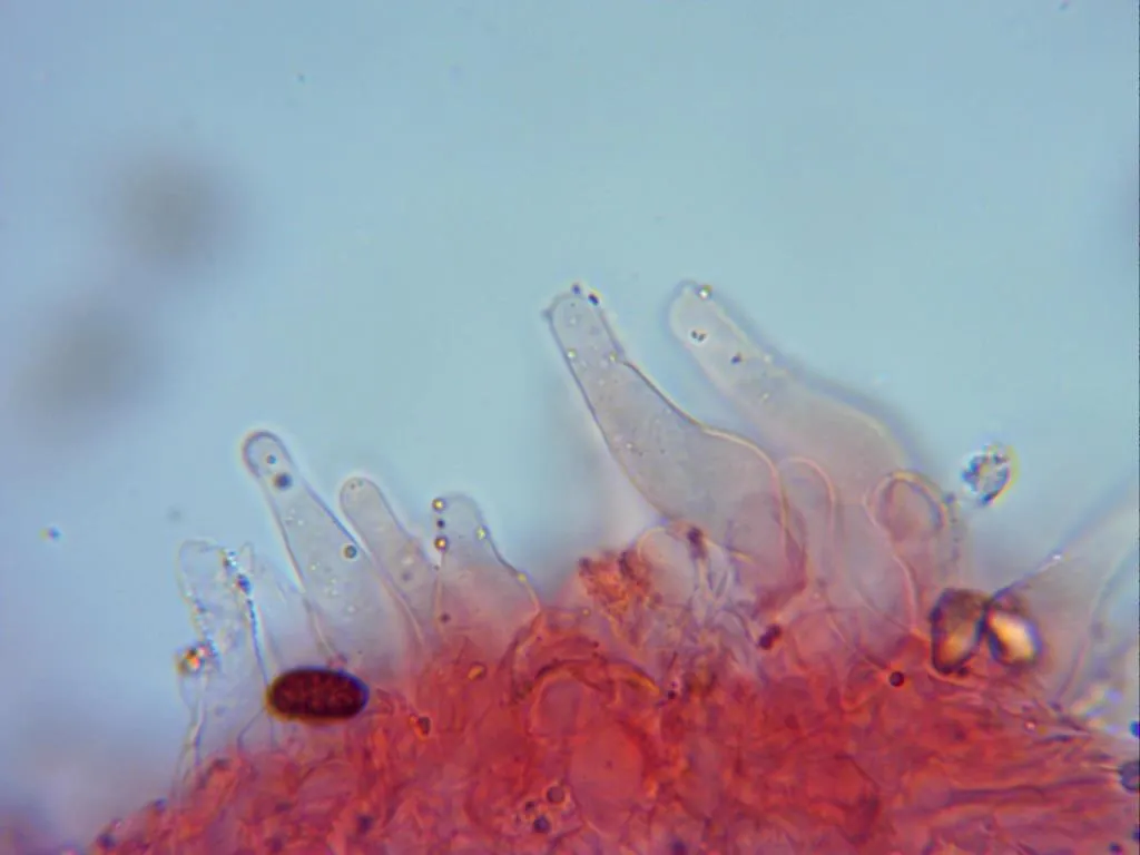
Caulocystidia present at the stipe apex, subcylindrical to lageniform.
Cheilocystidia abundant, fusiform to lageniform, 25 to 40 µm long and 8 to 11 µm wide.
Pleurocystidia scattered, 55 to 80 µm long and 11 to 16 µm wide.
Basidia clavate, 22 to 35 µm long, 11 to 15 µm wide, partly clamped, 4-spored.
Spores cylindrical-ellipsoidal, smooth, dark reddish-brown under the microscope, 9.5 to 12 µm long, 5 to 7 µm wide, with an inconspicuous eccentric apiculus at one pole and a conspicuous germ pore at the opposite pole.
Herbarium material available: JTP2799/25.
Geographic references
Map generated from global occurrence data from GBIF. GBIF.
Study measurements
Values in Microns (µm)Macroscopic gallery
.webp)
Microscopic gallery
Genetic / Phylogenetic Study
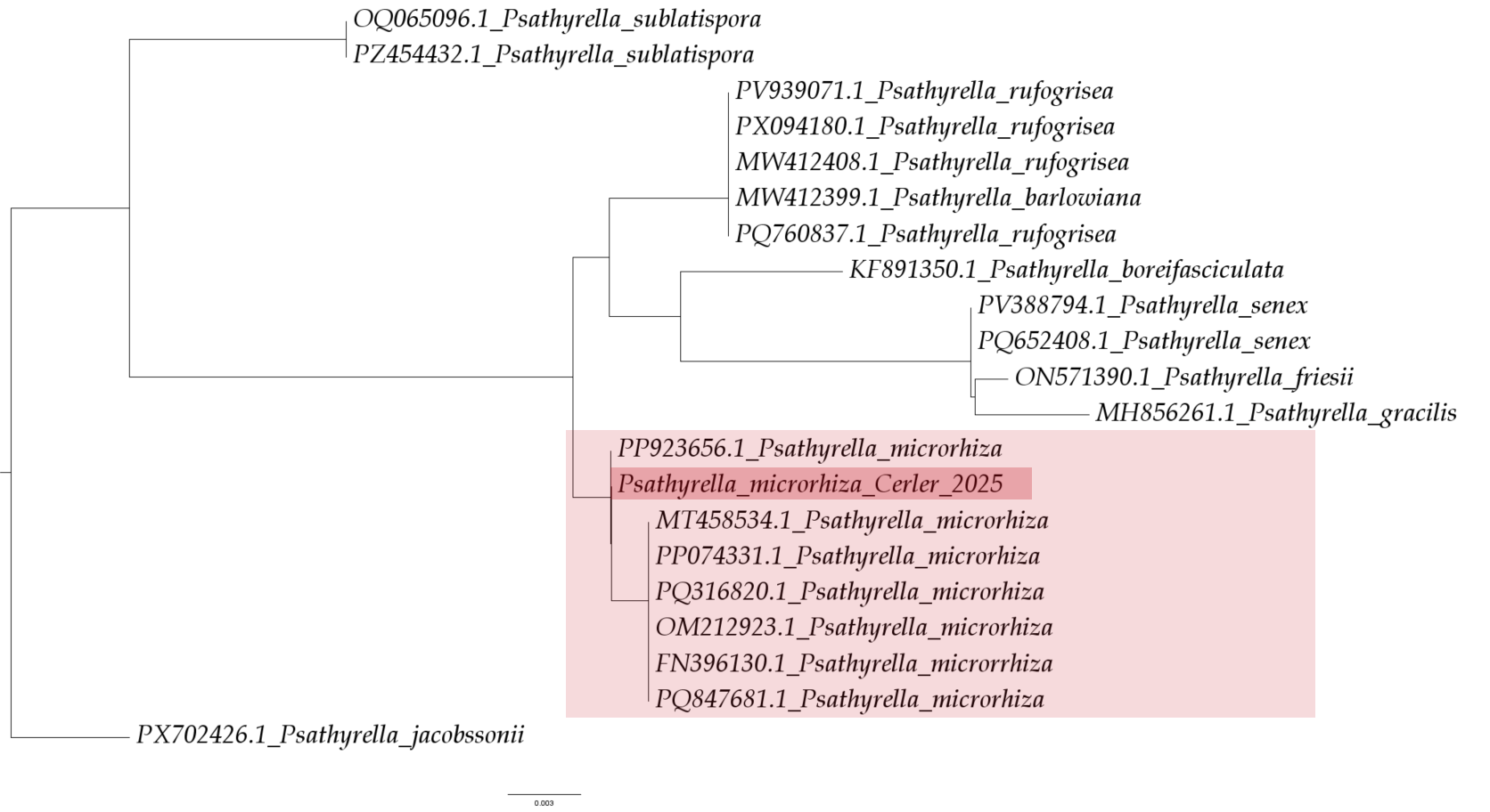
Phylogenetic Tree
Sequences and Data
Sequencing (Chromatogram)
Select a file to visualize the chromatogram