Psathyrella bipellis
(Quél.) A.H.Sm.
Study description
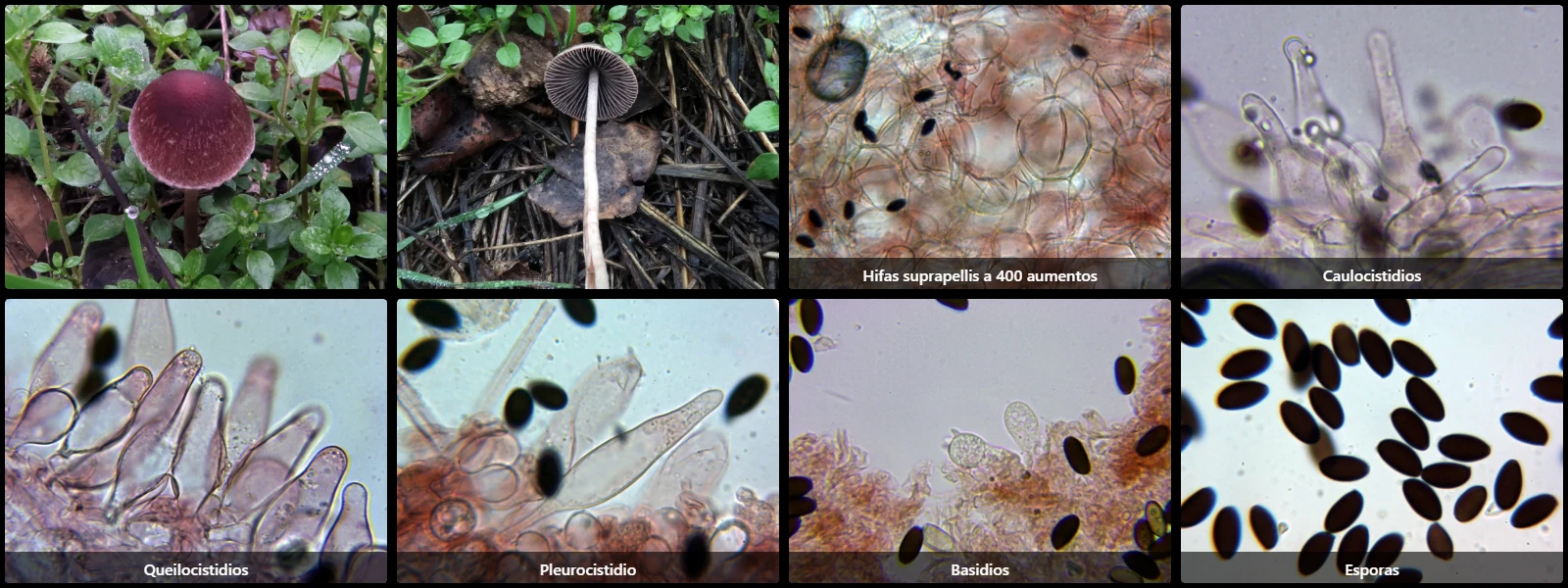
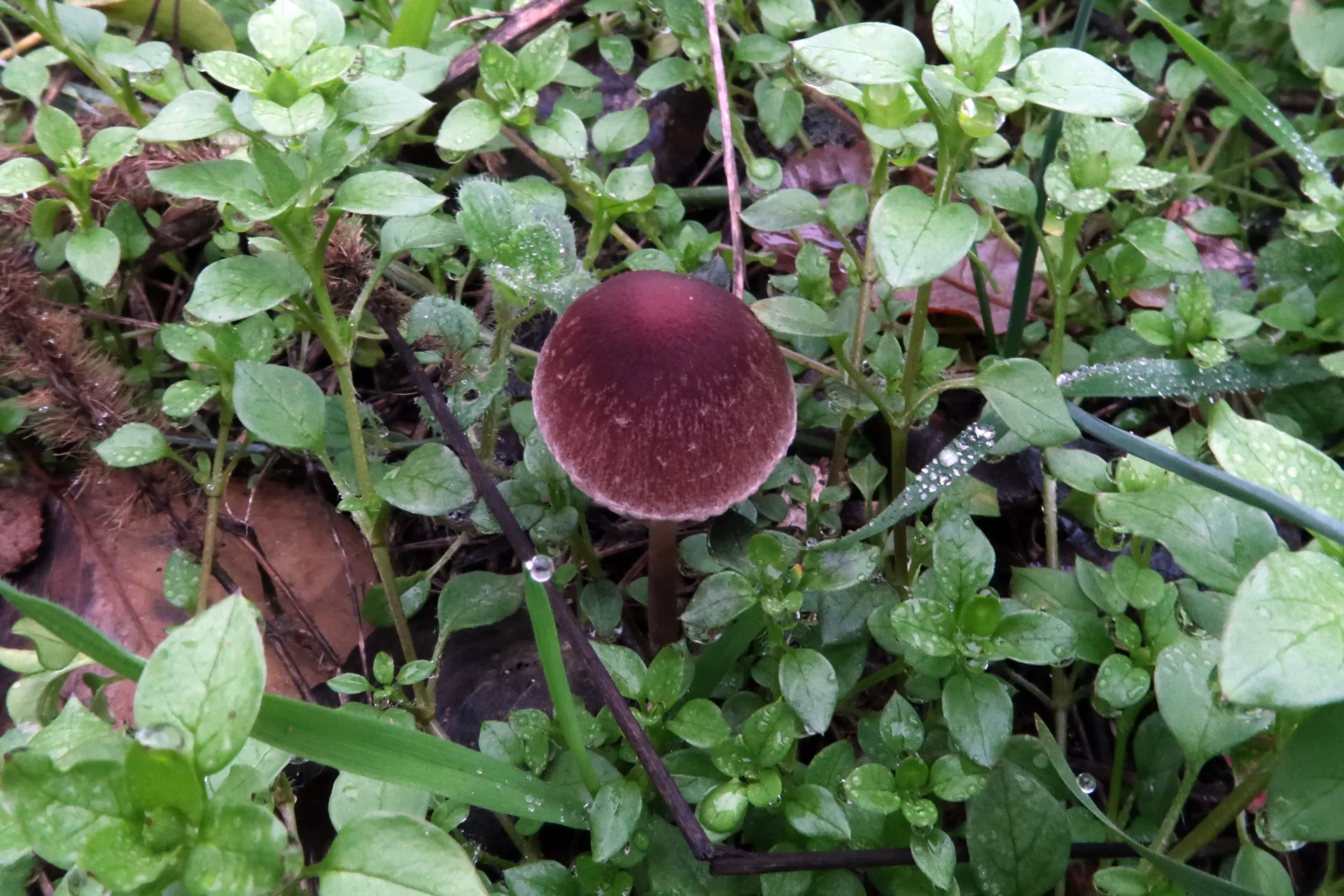
On woody debris on the ground or directly on decaying wood, with a convex to umbonate pileus 1.5 to 3.5 cm in diameter, reddish-brown, darker at the disc, with a whitish margin adorned with veil remnants, ascending, adnate lamellae, pinkish to dark grey, a cylindrical, hollow stipe, brownish-grey towards the base, fruity odour, and mild taste.
Pileipellis cellular, composed of subglobose to pyriform cells 20 to 50 µm in diameter, containing pale brownish intracellular pigment.
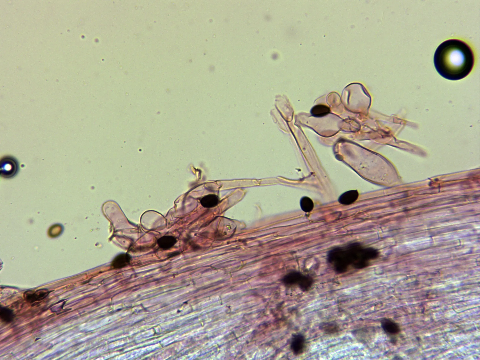
Caulocystidia subcylindrical, fusiform to lageniform, clustered in fascicles.
Cheilocystidia pyriform, clavate to lageniform, 35 to 50 µm long and 11 to 16 µm wide, often bearing small apical droplets.
Pleurocystidia fusiform to lageniform, 65 to 75 µm long and 12 to 15 µm wide.
Basidia clavate, 35 to 40 µm long, 13 to 16 µm wide, clampless at the base, 2- or 4-spored.
Spores ellipsoidal, dark brown under the microscope, 12 to 15 µm long, 7 to 8 µm wide, with a tiny eccentric hyaline apiculus at one end and a conspicuous central germ pore at the opposite pole.
Geographic references
Map generated from global occurrence data from GBIF. GBIF.
Study measurements
Values in Microns (µm)Macroscopic gallery
Microscopic gallery