Psathyrella arenosa
Örstadius & E.Larss.
Study description
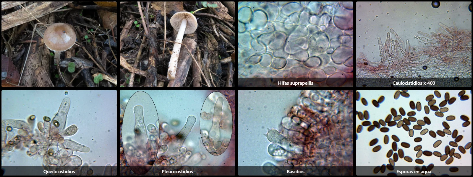
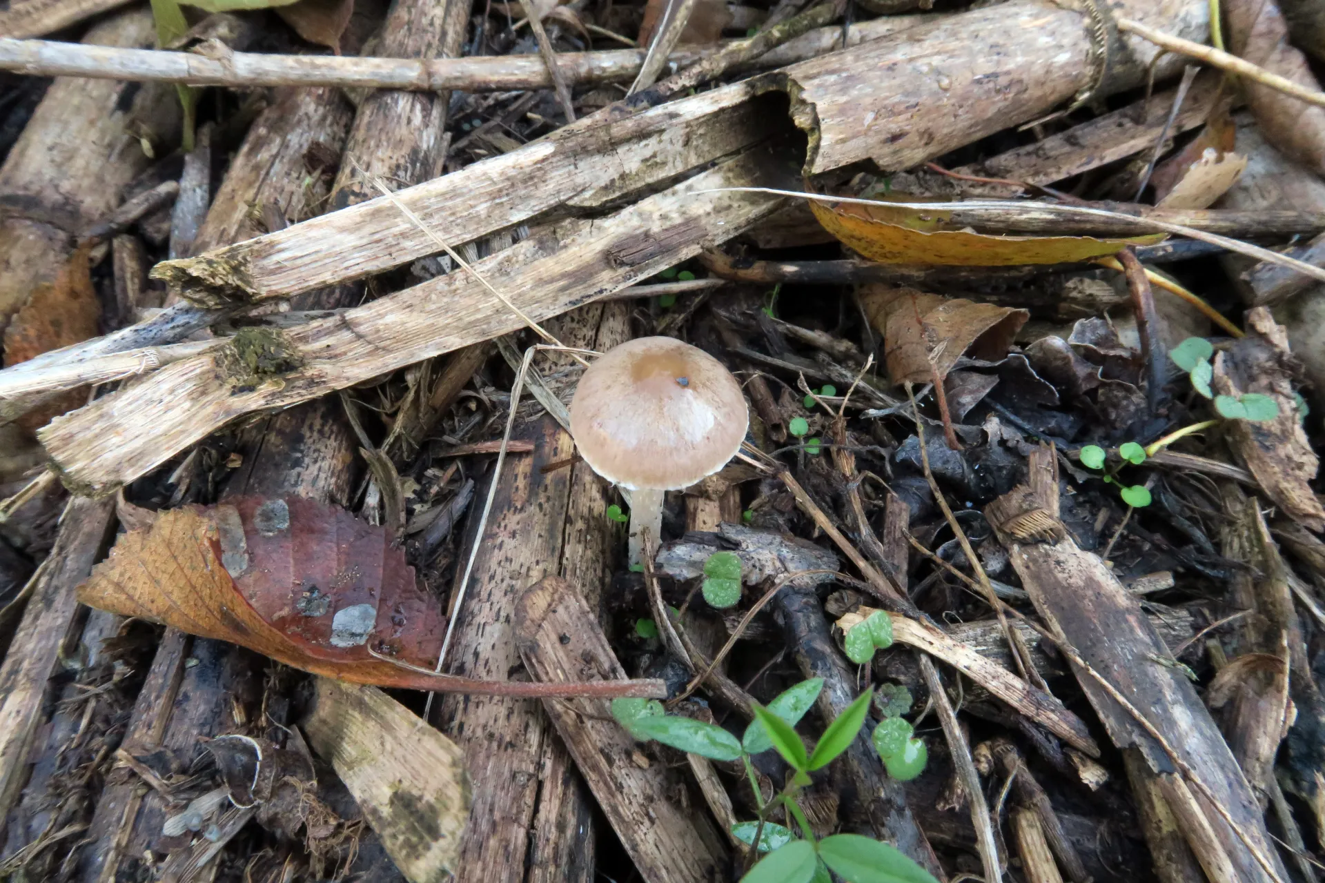
On calcareous soils among decaying debris of Phragmites, with a hemispherical to convex pileus 0.5 to 1.5 cm in diameter, yellowish-brown, radially fibrillose, white lamellae turning grey with purplish tints from sporulation, and a cylindrical stipe slightly enlarged at the base, pale brown.
Pileipellis cellular, composed of globose to pyriform cells 15 to 40 µm long and 12 to 30 µm wide, overlying interwoven filamentous hyphae 4 to 10 µm wide, septate with clamp connections.
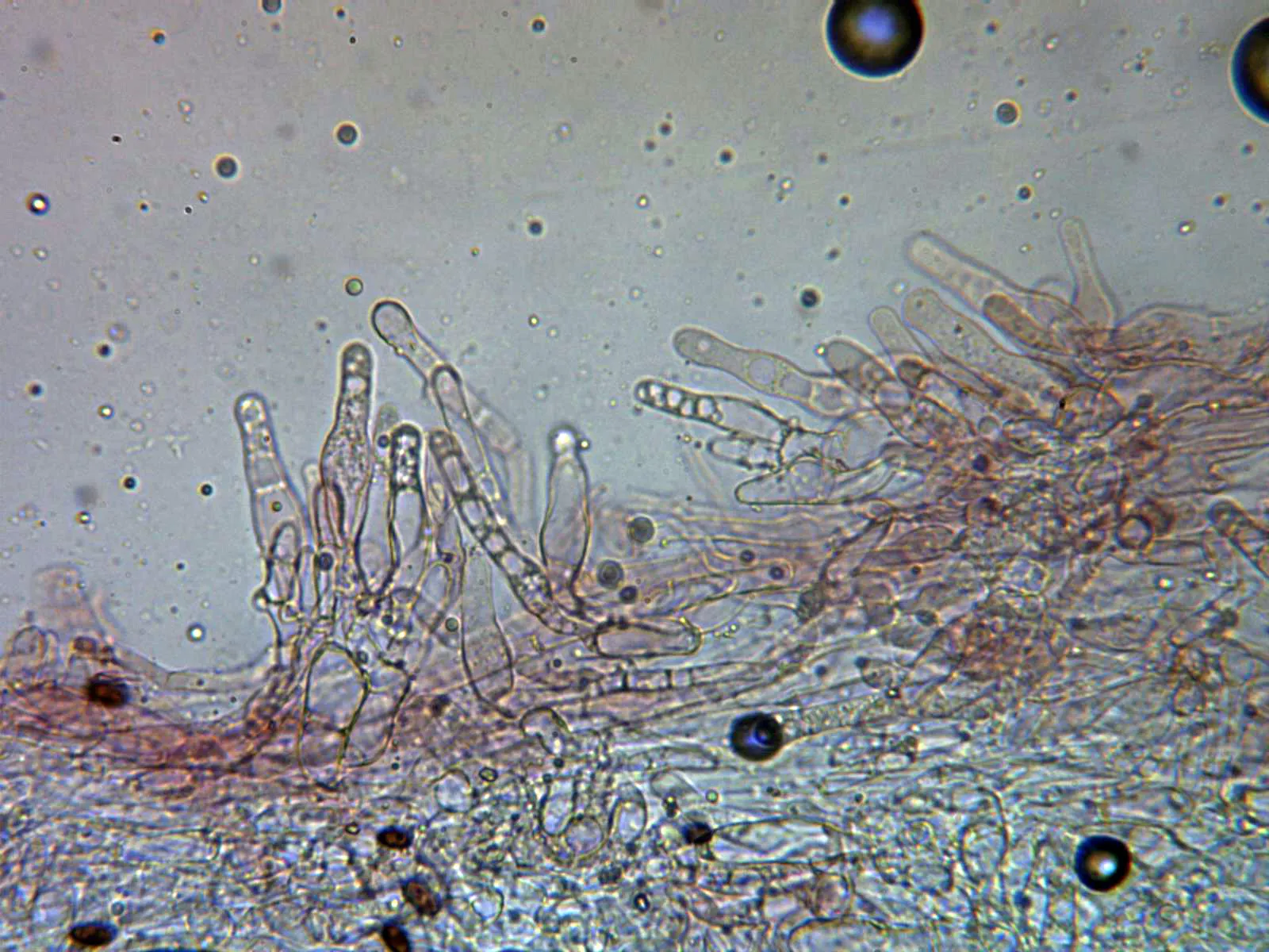
Caulocystidia present only at the stipe apex, subcylindrical to lageniform, sinuous, arranged in tufts.
Cheilocystidia subcylindrical, pyriform to lageniform, 22 to 40 µm long and 8 to 16 µm wide.
Pleurocystidia scattered, scarce, lageniform, 40 to 55 µm long and 10 to 13 µm wide.
Basidia clavate, 20 to 25 µm long, 7 to 9 µm wide, clampless at the base, 4-spored.
Spores cylindrical-ellipsoidal, smooth, thick-walled, brown under the microscope, 7 to 9 µm long, 4 to 5 µm wide, lacking an obvious apiculus, with a central germ pore at one pole, containing small guttules inside.
Geographic references
Map generated from global occurrence data from GBIF. GBIF.
Study measurements
Values in Microns (µm)Macroscopic gallery
Microscopic gallery