Pluteus thomsonii
(Berk. & Broome) Dennis
Study description
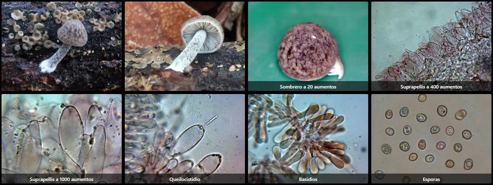
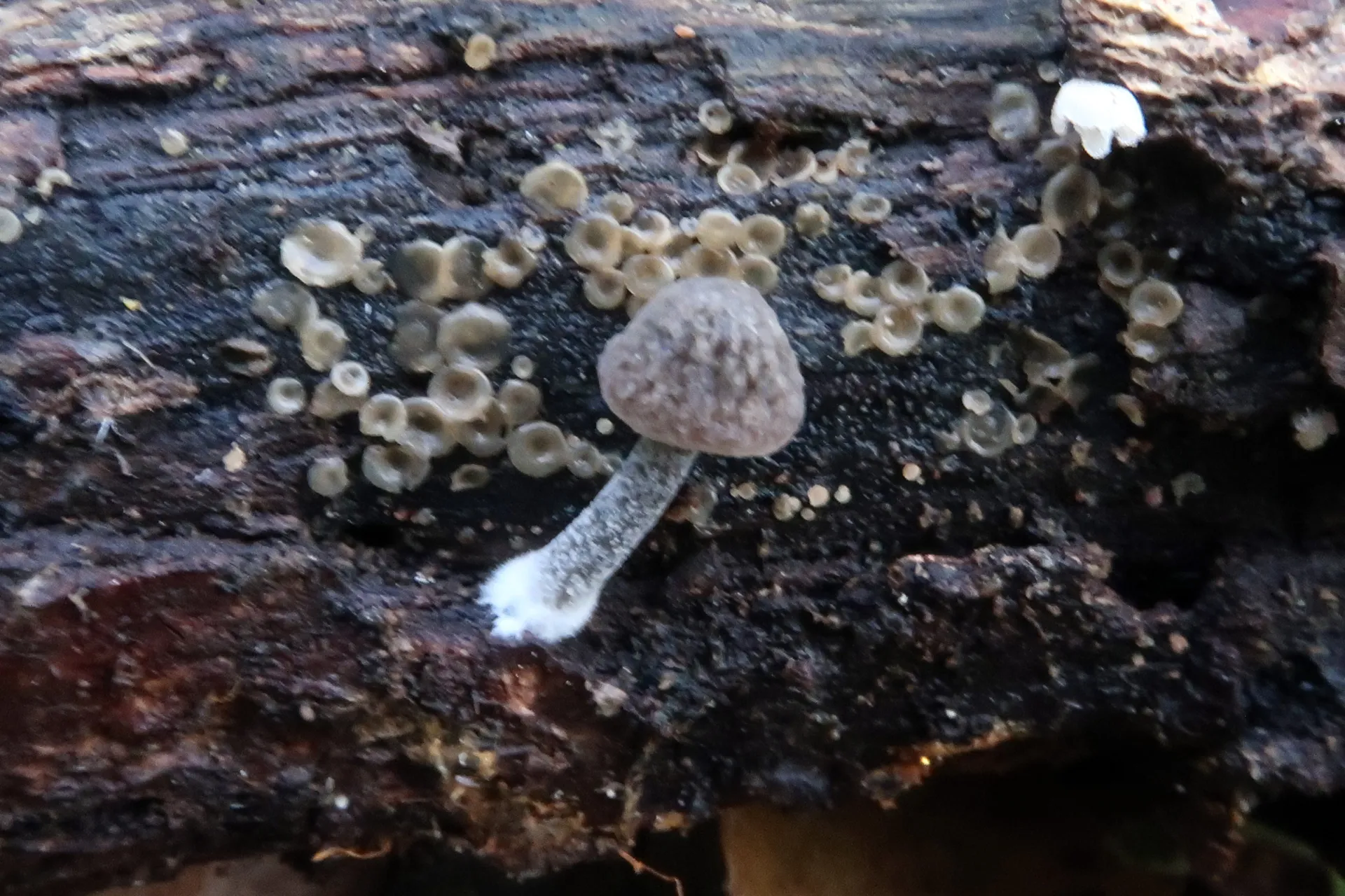
On decaying dead wood on the ground, with a convex to flattened pileus 1 to 3 cm across, dark brown, distinctly reticulate-venose surface, free white lamellae turning pink with development, a cylindrical, slightly enlarged greyish-white stipe, and mild, indistinct odour and taste.
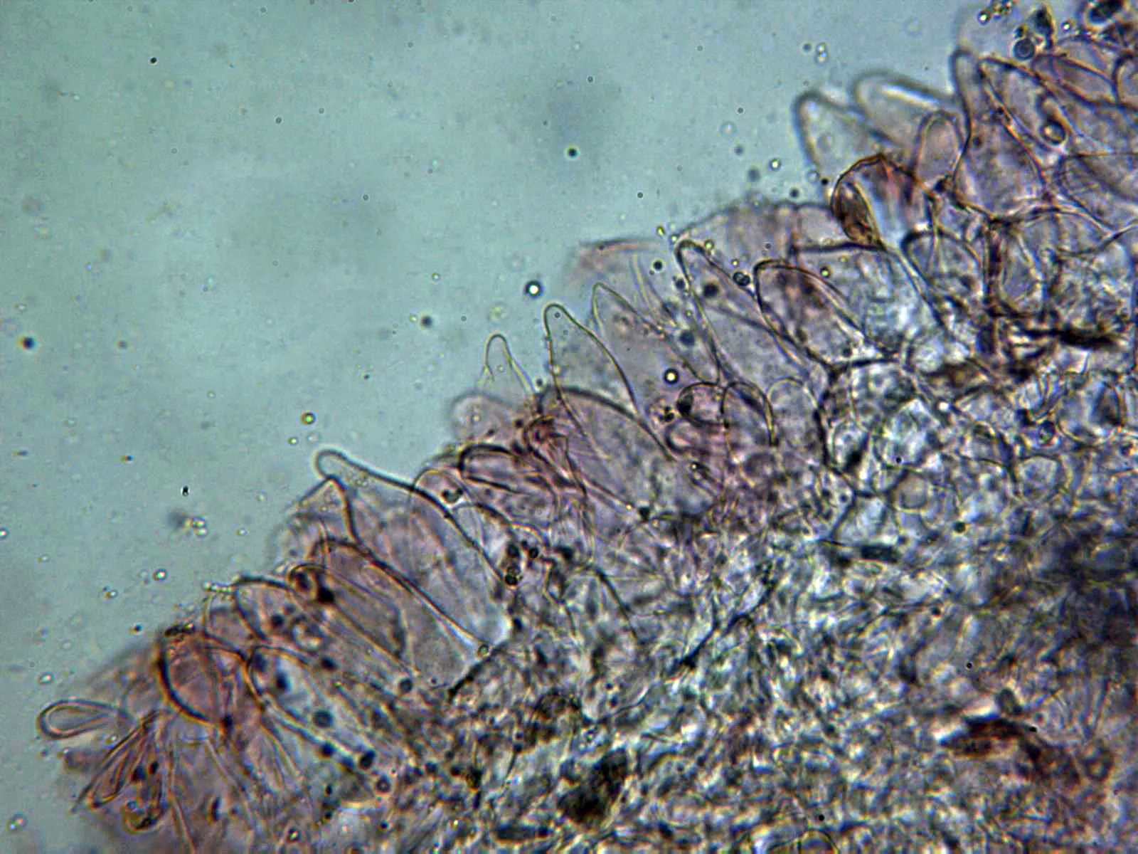
Pileipellis with emergent pileocystidia that are subcylindrical, clavate to fusiform, 50 to 80 µm long and 15 to 25 µm wide, filled with brown intracellular pigment.
Cheilocystidia fusiform, with an elongated apical rostrum/neck, 30 to 55 µm long and 10 to 16 µm wide.
Pleurocystidia not observed, if present very scarce and similar in morphology and size to cheilocystidia.
Basidia clavate, 20 to 30 µm long, 5 to 7 µm wide, simple-septate at the base, 4-spored.
Spores subglobose to ovoid, smooth, hyaline, moderately thick-walled, 6 to 8 µm long, 5 to 7 µm wide, with an inconspicuous hyaline apiculus, containing oil guttules, inamyloid in Melzer's reagent.
Geographic references
Map generated from global occurrence data from GBIF. GBIF.
Study measurements
Values in Microns (µm)Macroscopic gallery
Microscopic gallery