Pluteus romellii
(Britzelm.) Lapl.
Study description
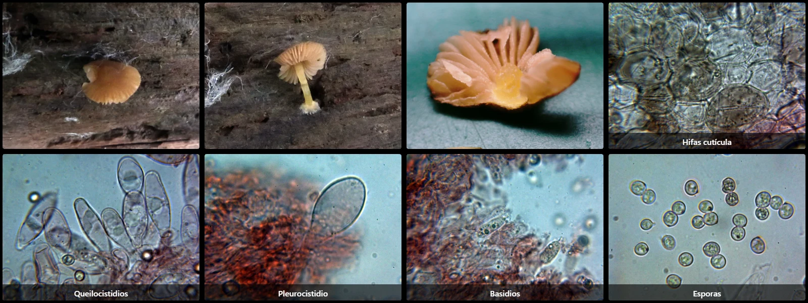
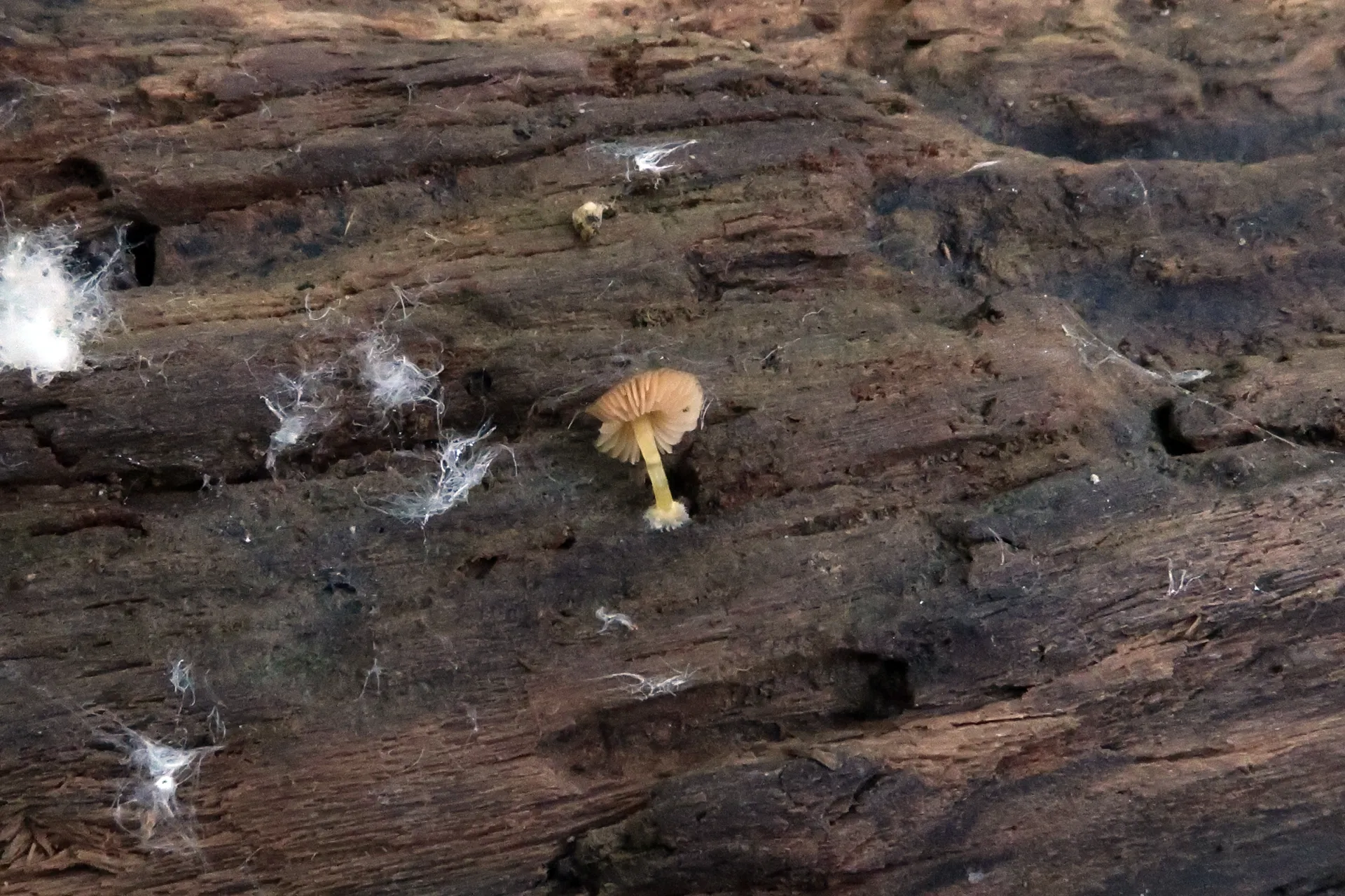
On stumps or decaying fallen wood in deciduous or mixed woodlands, with a convex to flattened pileus 1.5 to 3 cm in diameter, yellowish-brown with a slightly darker centre, free white lamellae turning pink from sporulation, a cylindrical, smooth, lustrous bright yellow stipe, practically odourless, and with a slightly raphanoid taste.
Pileipellis cellular (a hymeniderm), composed of subglobose to pyriform cells 30 to 60 µm long and 20 to 50 µm wide, filled with brown intracellular pigment, clampless.
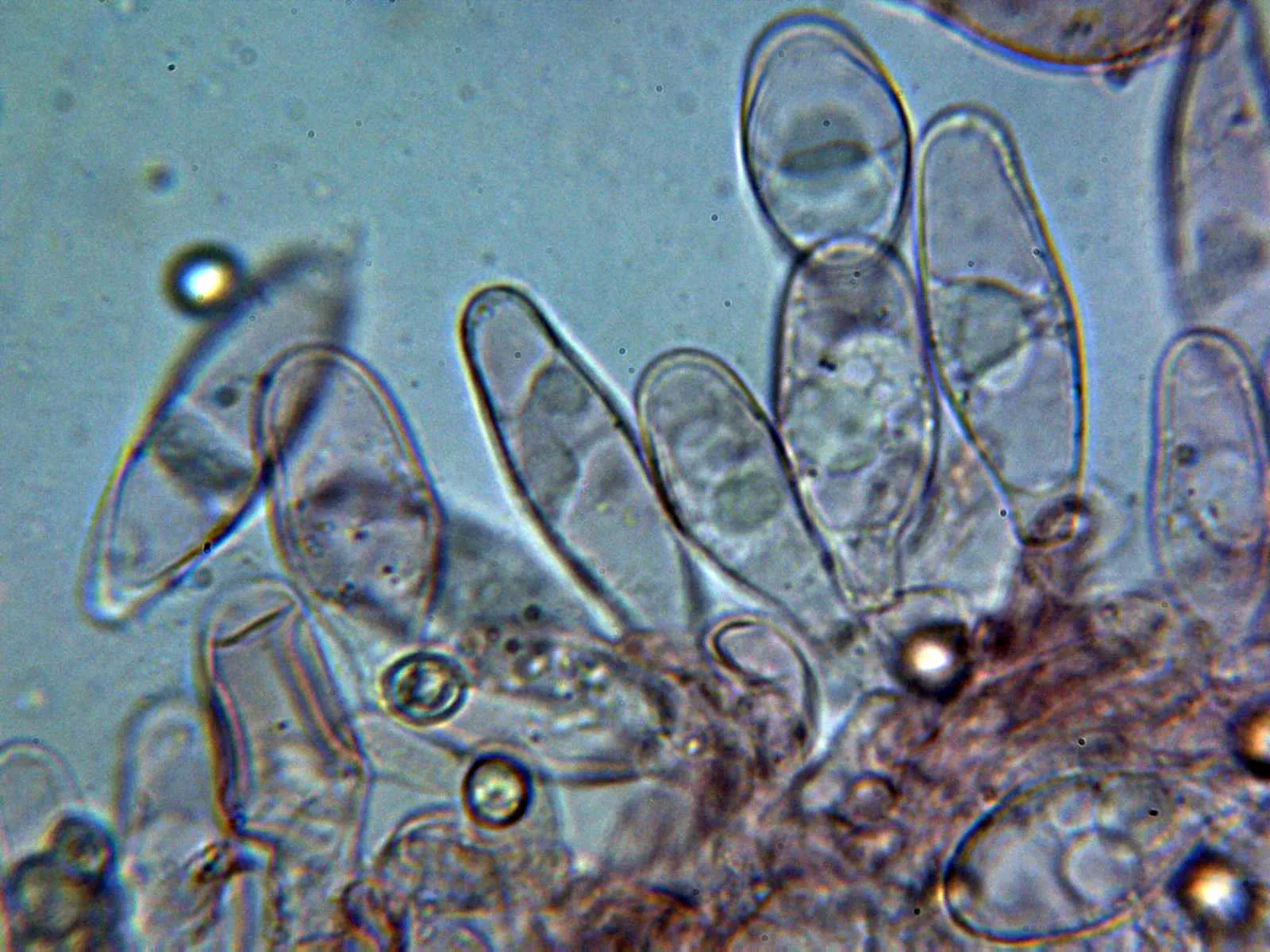
Cheilocystidia subcylindrical, clavate to fusiform, 30 to 60 µm long and 15 to 30 µm wide.
Pleurocystidia scarce, clavate to ventricose, 45 to 60 µm long and 25 to 35 µm wide.
Basidia cylindrical, 20 to 30 µm long, 8 to 10 µm wide, simple-septate at the base, 2- or 4-spored.
Spores subglobose to ovoid, smooth, slightly thick-walled, pinkish-grey under the microscope, 5.5 to 7 µm long, 4.5 to 6.5 µm wide, with an inconspicuous apiculus, containing oil droplets, inamyloid in Melzer's reagent.
Geographic references
Map generated from global occurrence data from GBIF. GBIF.
Study measurements
Values in Microns (µm)Macroscopic gallery
Microscopic gallery