Pluteus phlebophorus
(Ditmar) P.Kumm.
Study description
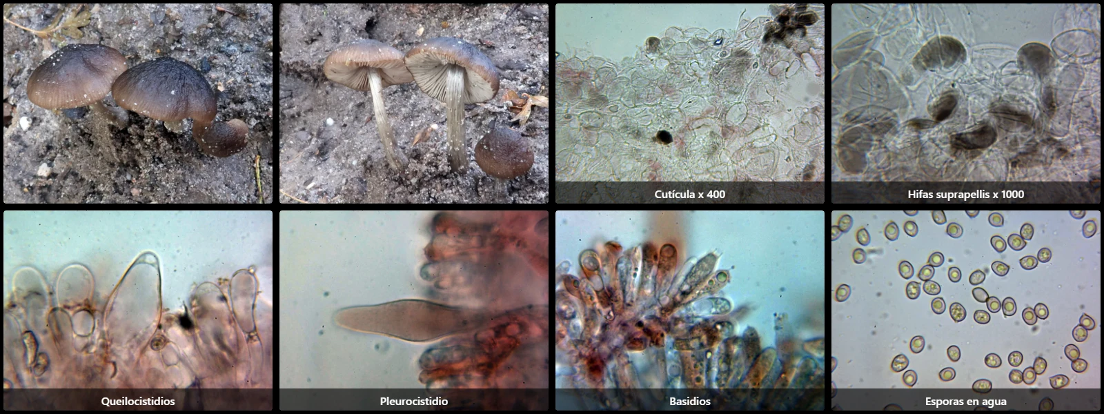
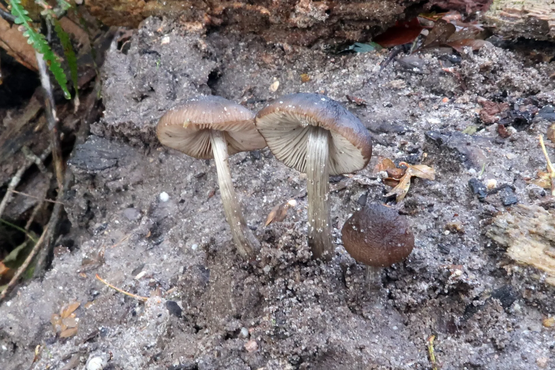
In forest clearings on heavily decayed wood, with a convex to flattened pileus 2 to 4.5 cm in diameter, reddish-brown, darker at the disc, radially venose-wrinkled, free lamellae, initially white then pink from sporulation, a cylindrical greyish-ochre stipe covered with whitish longitudinal fibrils, raphanoid odour, and insipid taste.
Pileipellis cellular (a hymeniderm), composed of globose to pyriform cells 35 to 50 µm long and 20 to 35 µm wide, clampless, containing abundant brown intracellular pigment.
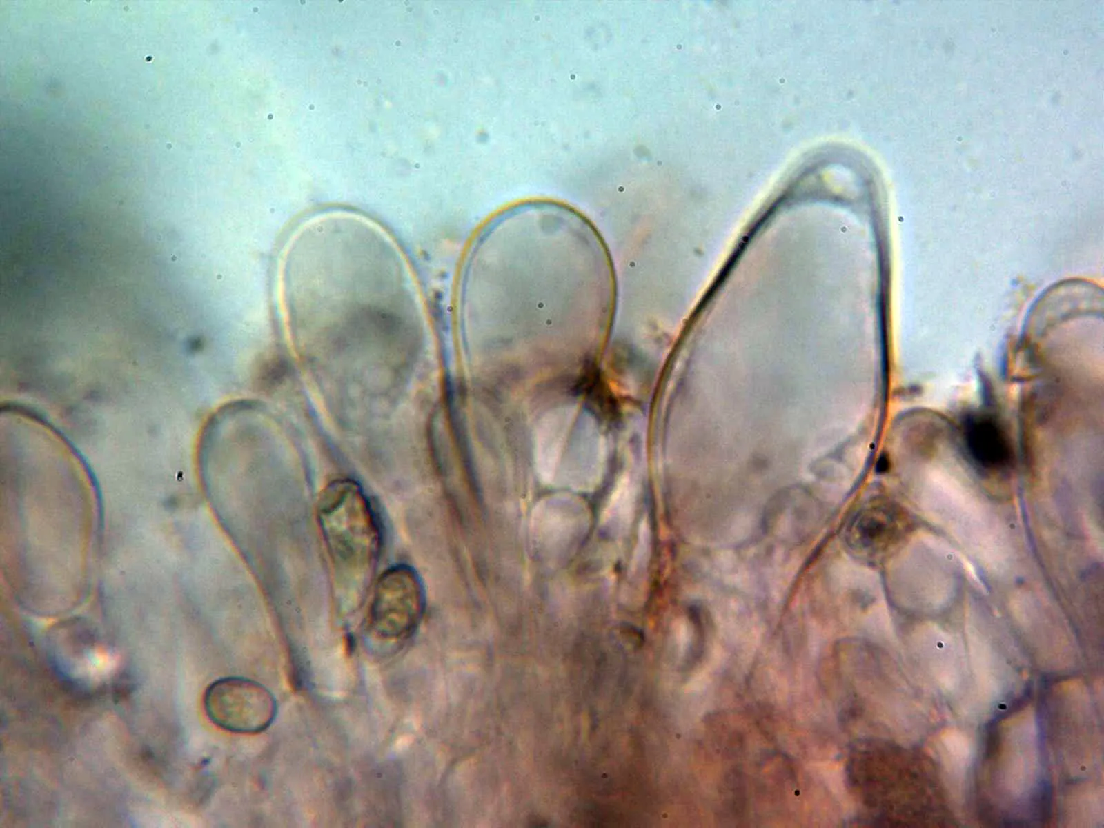
Cheilocystidia fusiform to clavate, 50 to 75 µm long and 15 to 30 µm wide.
Pleurocystidia fusiform to lageniform, 60 to 90 µm long and 15 to 20 µm wide.
Basidia cylindrical, slightly ventricose, 25 to 35 µm long, 8 to 11 µm wide, simple-septate at the base, 4-spored.
Spores subglobose to ellipsoidal, smooth, moderately thick-walled, pinkish-grey under the microscope, 6.5 to 8.5 µm long, 5 to 6.5 µm wide, with a hyaline apiculus at one end, containing guttules, inamyloid in Melzer's reagent.
Geographic references
Map generated from global occurrence data from GBIF. GBIF.
Study measurements
Values in Microns (µm)Macroscopic gallery
Microscopic gallery