Piloderma byssinum
(P.Karst.) Jülich
Study description
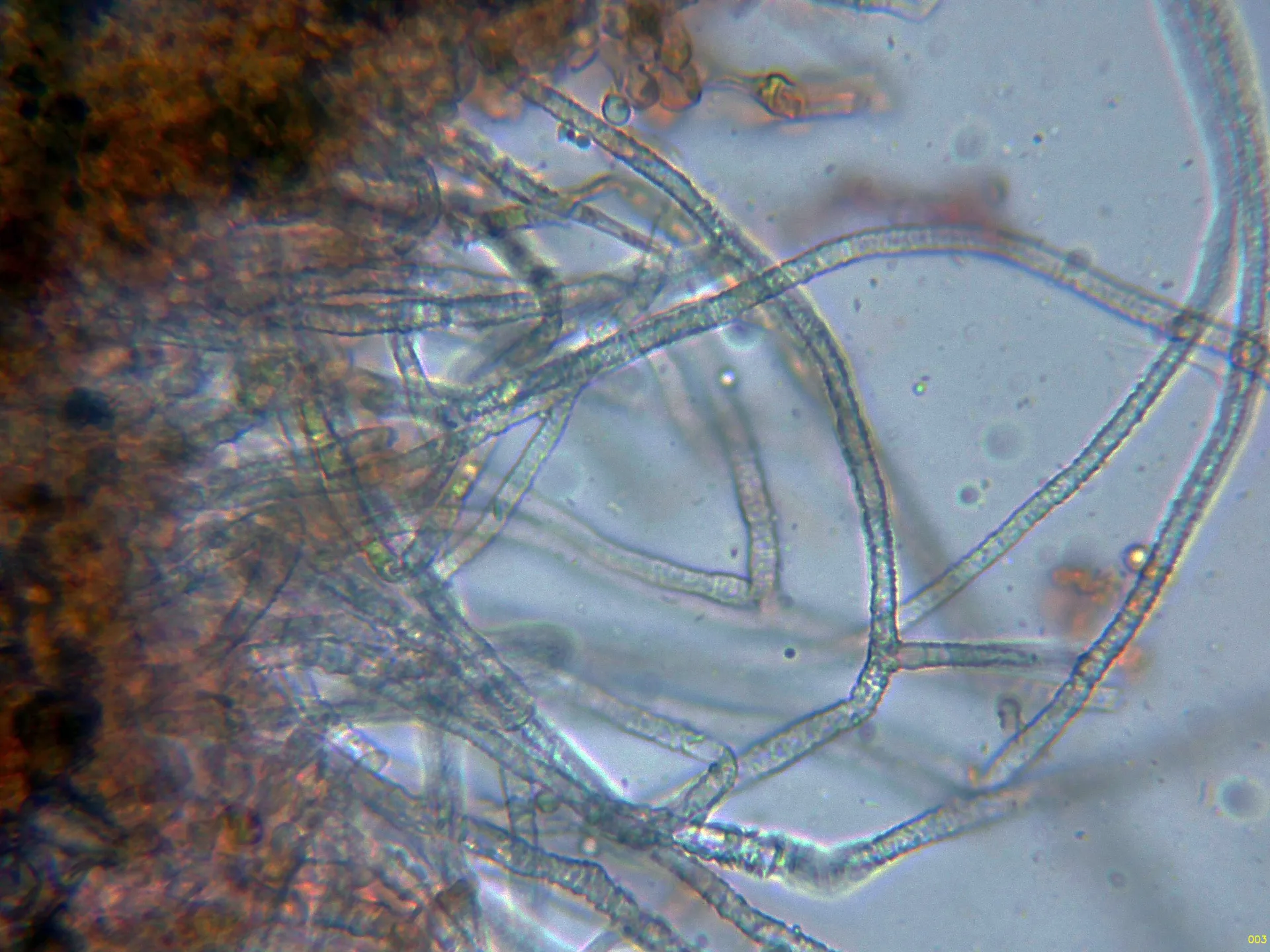
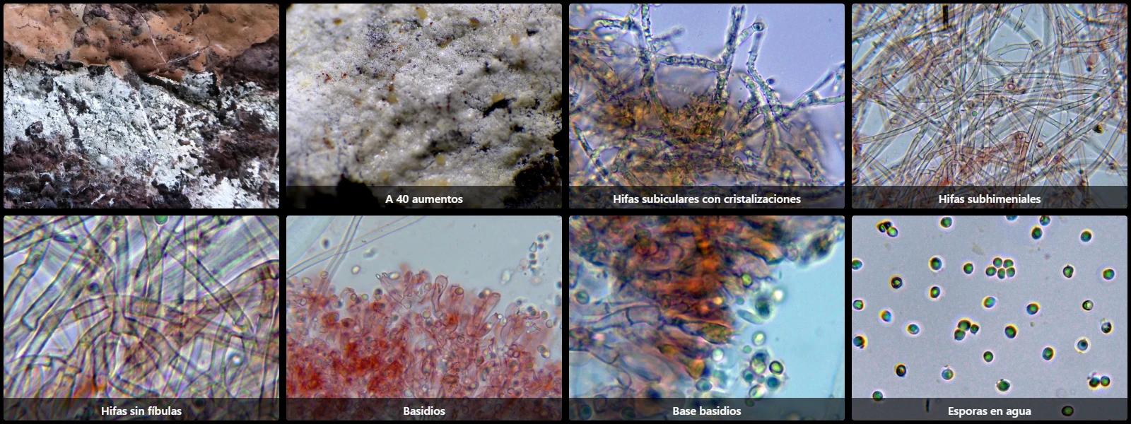
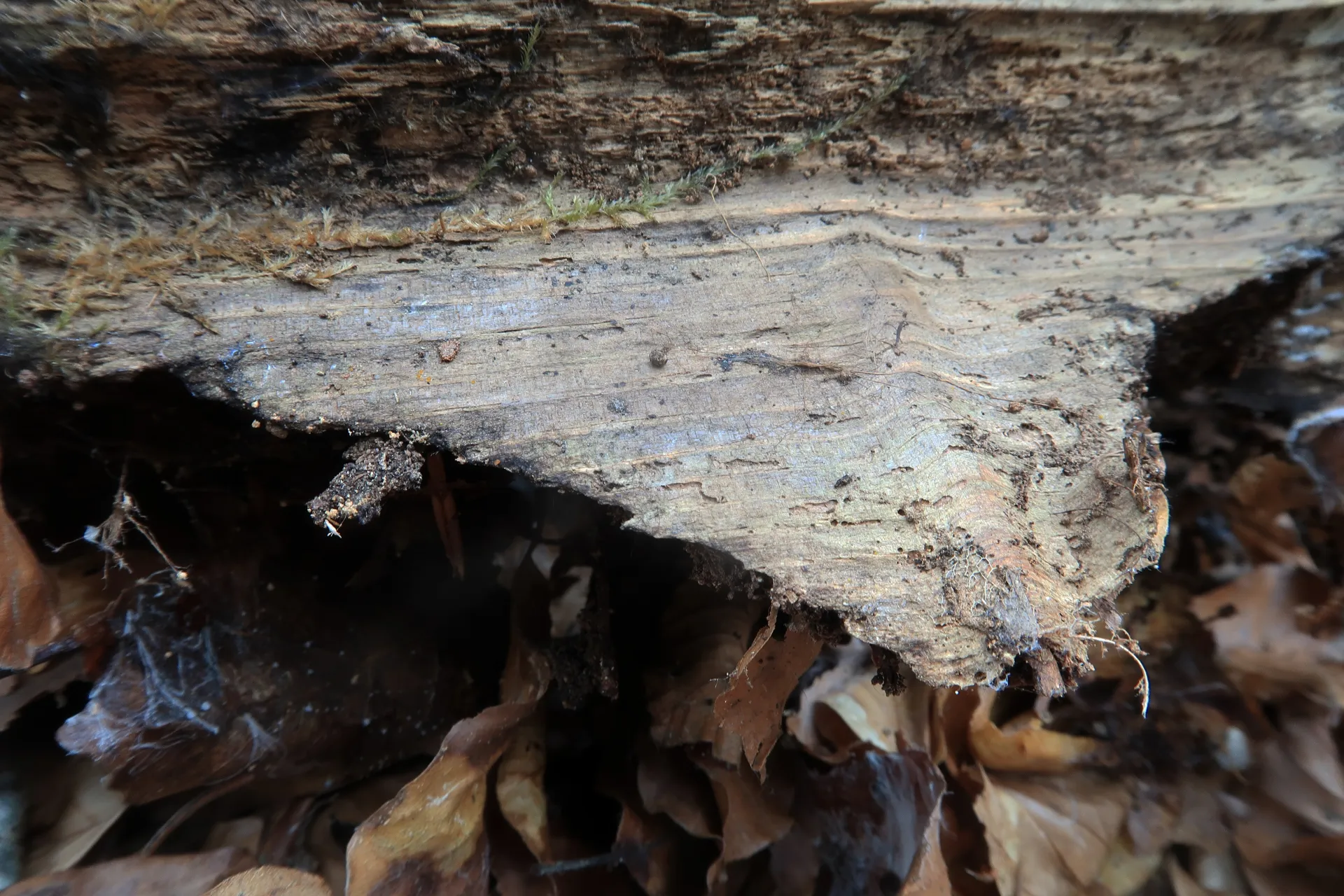
On wood in an advanced state of decay, of both conifers and broadleaved trees, with white, byssoid basidiomata only 0.1 to 0.3 mm thick, easily detached from the substrate, with abundant white rhizomorphs radiating at the growth margins.
Hyphal system monomitic; subicular hyphae thick-walled, 2.5 to 4 µm wide, densely covered with acicular crystals; subhymenial hyphae hyaline, smooth, thin-walled, 2 to 3 µm wide; both septate with simple septa (lacking clamps).
Cystidia and other sterile hymenial elements absent.
Basidia clavate, 10 to 20 µm long, 4 to 6 µm wide, simple-septate at the base, 4-spored.
Spores subglobose to ovoid, thick- and smooth-walled, hyaline, 3 to 4 µm long, 2.5 to 3.5 µm wide, with an inconspicuous apiculus at one pole, containing a prominent oil droplet, inamyloid in Melzer's reagent.
Herbarium material available: JTP2087/24.
Geographic references
Map generated from global occurrence data from GBIF. GBIF.
Study measurements
Values in Microns (µm)Macroscopic gallery
Microscopic gallery