


Phlegmacium balteatocumatile
(Rob.Henry ex P.D.Orton) Niskanen & Liimat.
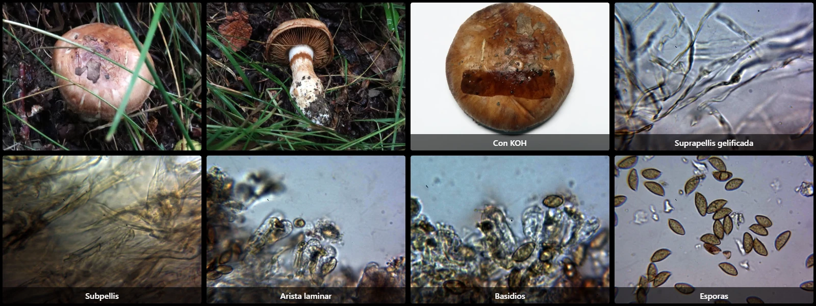
Study description
Suprapellis composed of slender hyphae widely separated by gelatinization and bearing clamp connections. Underneath, ochraceous pigmented and interwoven hyphae.
Lamellar edge without differentiated elements from basidia and basidioles.
Basidia cylindrical, up to 38 µm long, some with yellowish intracellular pigment, 4-spored.
Spores amygdaliform, thick-walled, with a lateral apiculus, yellowish-brown under the microscope with darker brown warts, with only a very weakly dextrinoid reaction in Melzer's reagent.
Geographic references
Map generated from global occurrence data from GBIF. GBIF.
Study measurements
Values in Microns (µm)Macroscopic gallery
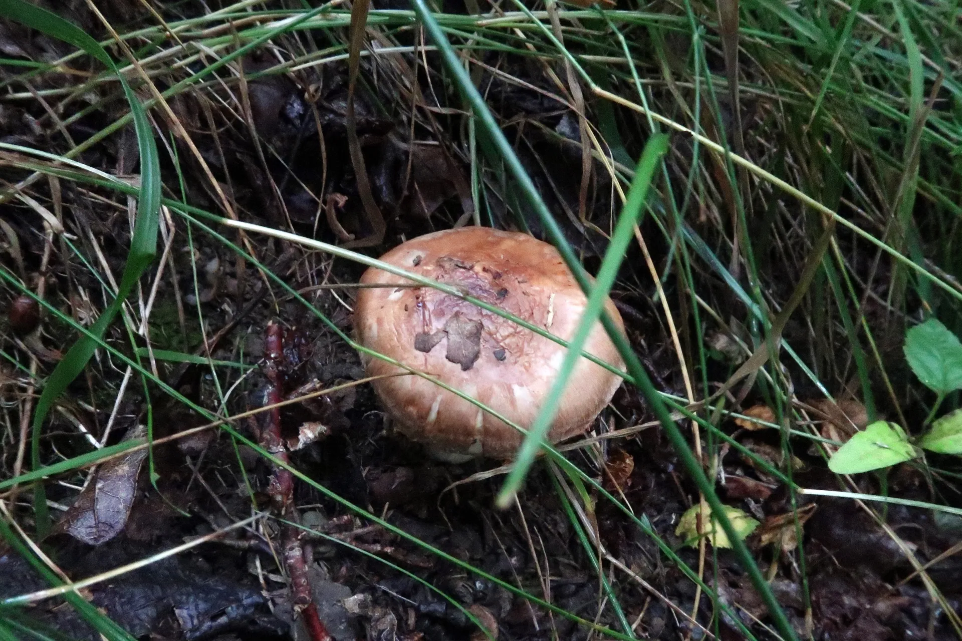
1 / 4Phlegmacium balteatocumatile - 1 - IMG_1444
Microscopic gallery
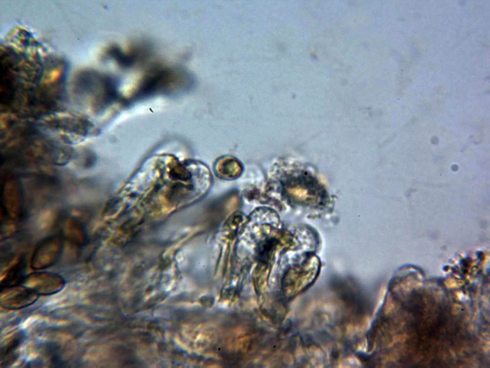
1 / 11Phlegmacium balteatocumatile - 10 - Arista laminar