Phlebia radiata
Fr.
Study description
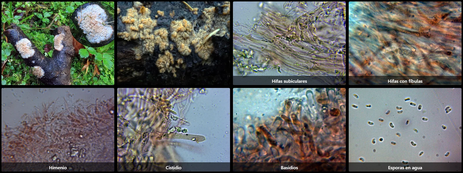
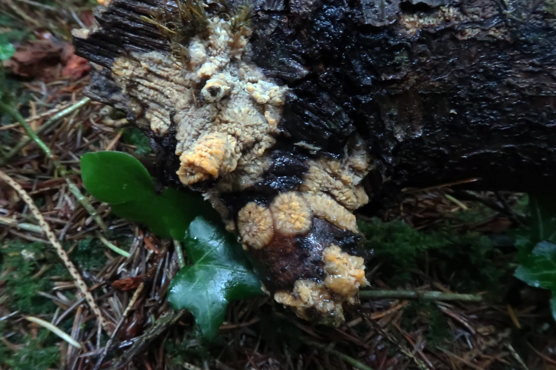
On decaying dead wood or bark on the ground, with completely resupinate basidiomata tightly adnate to the substrate, orange to pinkish, with a wrinkled to merulioid/tubercular hymenium arranged in radiating folds, a whitish growth margin, gelatinous when fresh and drying hard and corneous.
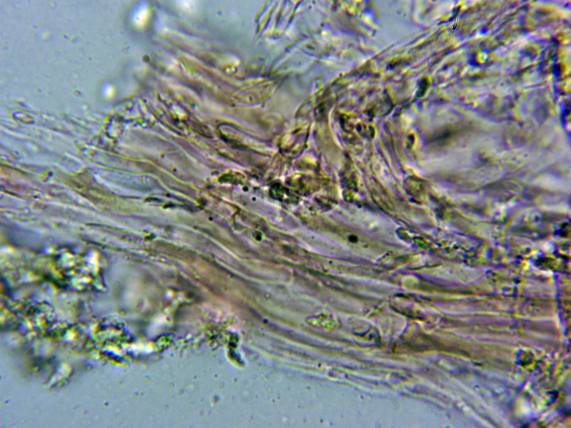
Hyphal system monomitic; subicular hyphae cylindrical, thin-walled, arranged more or less parallel, 2 to 4 µm wide, with faint brownish intracellular pigment; subhymenial hyphae hyaline, 1.5 to 3 µm wide; both septate with clamp connections.
Leptocystidia scattered, cylindrical to fusiform, deeply embedded in the hymenium and sometimes difficult to locate, hyaline and thin-walled.
Basidia cylindrical-clavate, 25 to 30 µm long, 3 to 4 µm wide, clamped at the base, 4-spored.
Spores cylindrical to allantoid, smooth, hyaline, thin-walled, 3.5 to 4.5 µm long, 1.5 to 2 µm wide, containing two oil drops located at opposite poles, inamyloid in Melzer's reagent.
Geographic references
Map generated from global occurrence data from GBIF. GBIF.
Study measurements
Values in Microns (µm)Macroscopic gallery
Microscopic gallery