Perenniporia rosmarini
A.David & Malençon
Study description
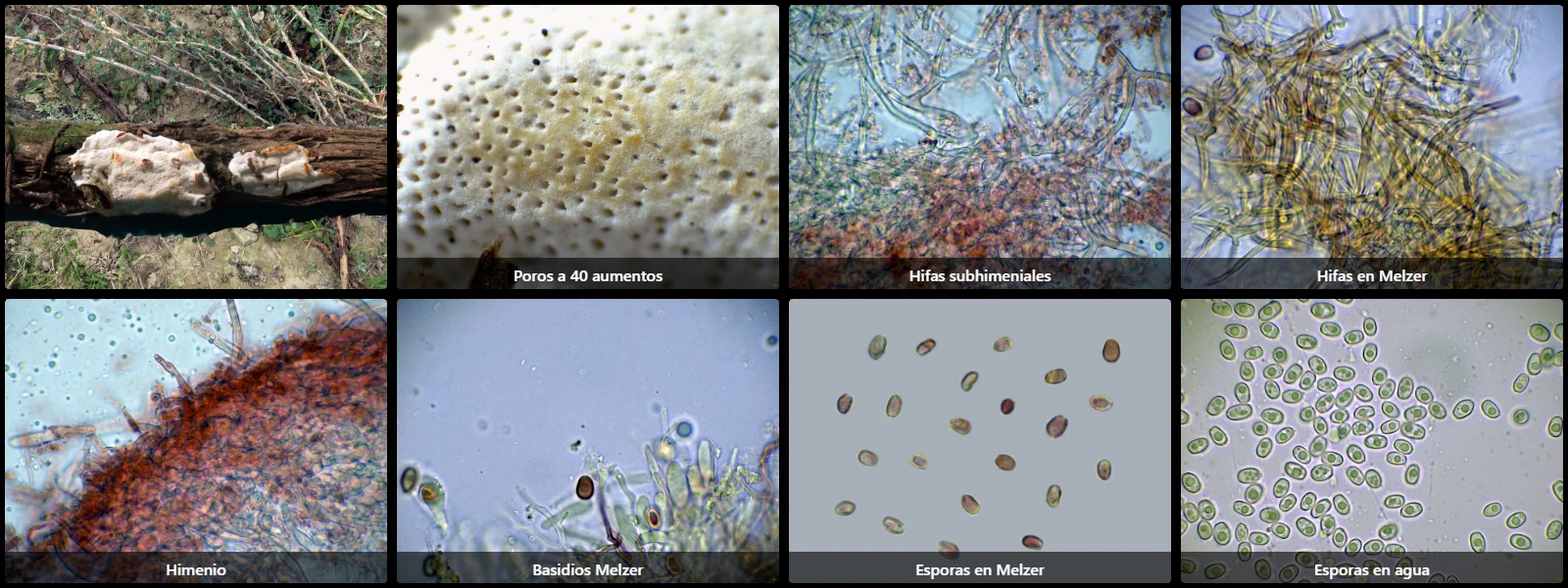
On dead decaying woody stems of Salvia rosmarinus on the ground, with a resupinate basidiocarp up to 15 cm long, white poroid surface with 6 to 7 pores per mm, a thin subiculum up to 1 mm thick, and a tube layer up to 2 mm deep.
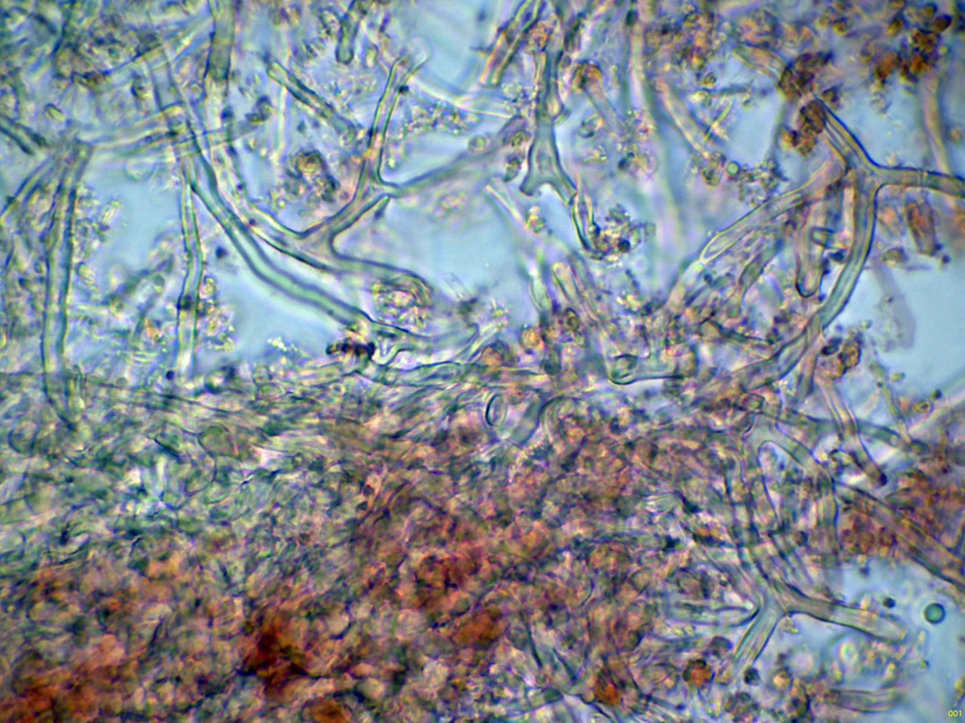
Hyphal system trimitic: skeletal hyphae thick-walled, aseptate, scarcely branched, 2 to 3 µm wide; generative hyphae thin-walled, 2 to 3 µm wide, septate with clamp connections; binding hyphae thick-walled, clampless, profusely branched.
Differentiated cystidia absent, but some fusiform cystidioles of similar dimensions to the basidia are present.
Basidia clavate, 20 to 25 µm long, 8 to 10 µm wide, clamped at the base, 4-spored.
Spores ellipsoidal, thick-walled, smooth, hyaline, 6.5 to 8 µm long, 5 to 6.5 µm wide, with a slightly eccentric apiculus at one pole, containing one or several guttules inside, dextrinoid in Melzer's reagent.
Herbarium material available: JTP2700/25.
Geographic references
Map generated from global occurrence data from GBIF. GBIF.
Study measurements
Values in Microns (µm)Macroscopic gallery
Microscopic gallery