Passalora acanthicola
(Hansf.) U.Braun & Crous
Study description
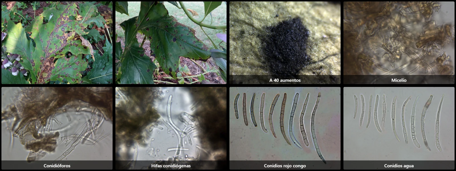
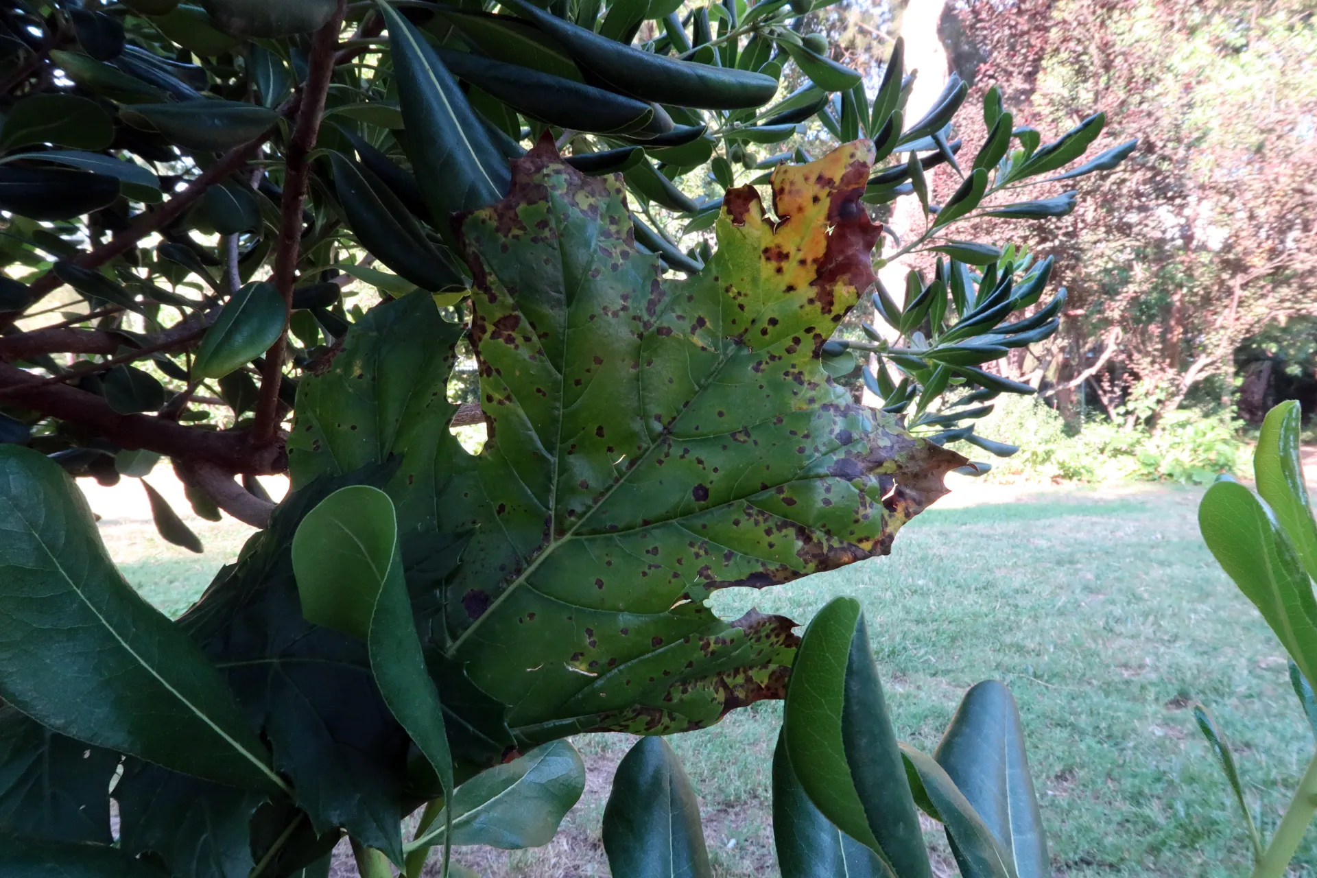
Formerly known as Cercospora acanthicola, documented in a public park in Barcelona on leaves of Acanthus mollis, causing necrotic foliar lesions (leaf spots) that are initially yellowish and darken to deep brown with age.
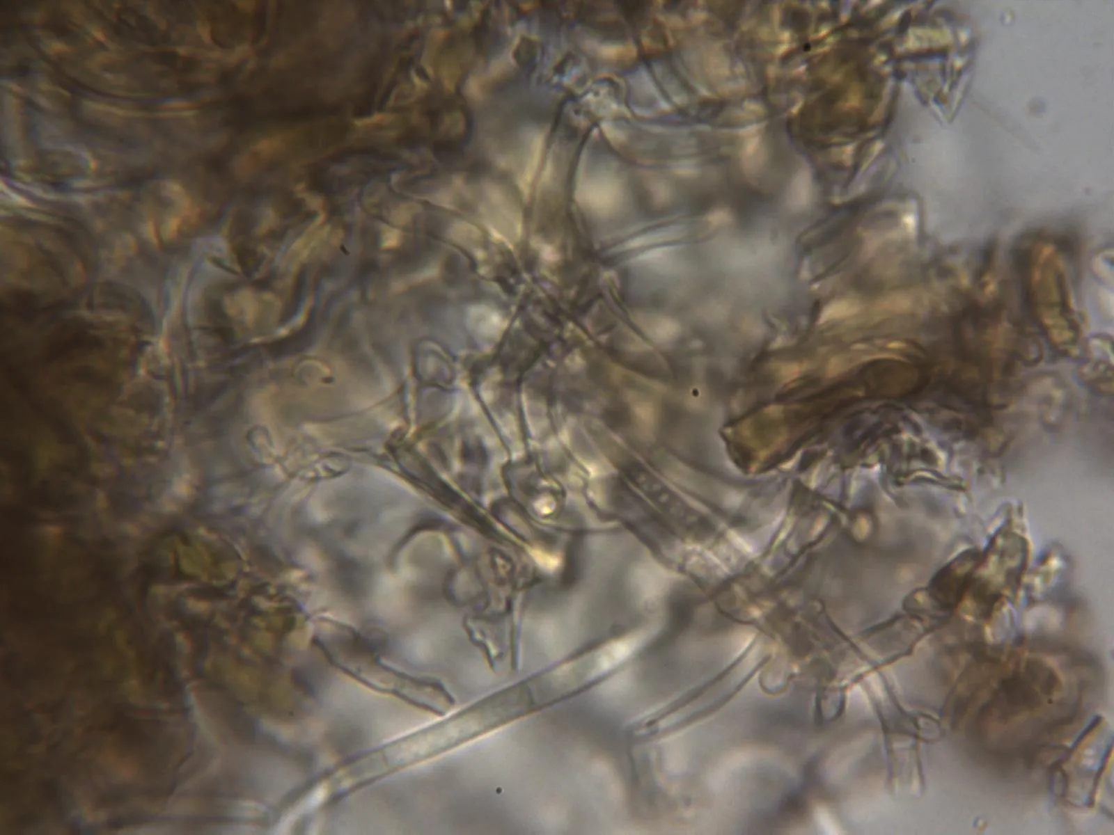
Without distinct pycnidia or stromata; intracellular mycelium composed of septate, branched hyphae 3–5 µm wide, hyaline to pale brown, thin- to slightly thick-walled.
Conidiophores fasciculate (grouped in bundles), measuring 150–230 µm long by 4–6 µm wide, straight to flexuous, bearing apical conidiogenous loci (conidial scars) 1.5–2.5 µm in diameter.
Conidia subcylindrical, straight or slightly curved at one end, with one obtuse and one slightly tapered end, smooth, hyaline, thin-walled, measuring 23–73 µm long by 2.5–3.5 µm wide, with 1 to 7 transverse septa.
Geographic references
Map generated from global occurrence data from GBIF. GBIF.
Study measurements
Values in Microns (µm)Macroscopic gallery
Microscopic gallery