Mucor racemosus
Fresen.
Study description
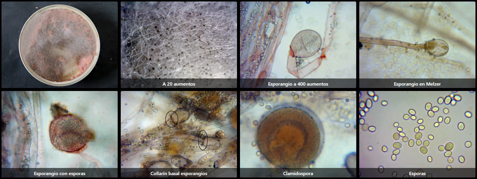
Species colonising a wide variety of foodstuffs, such as fruits, dairy products, and all types of processed food; the specimen studied here occurred on a spoiled tin of pâté, appearing as a cottony mass of abundant interwoven fine hyphae, hyaline and bearing numerous terminal minute black dots corresponding to sporangia.
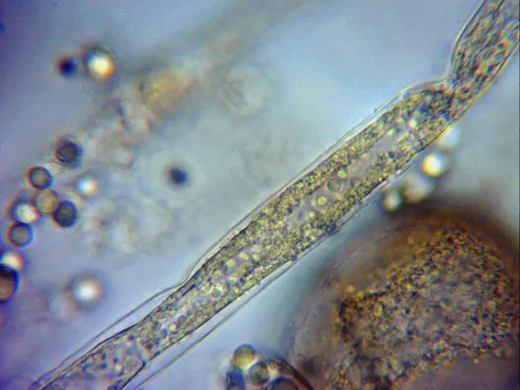
Mycelial hyphae coarse, hyaline, 5–15 µm wide, non-septate (coenocytic).
Sporangia globose, variable in size (30–70 µm in diameter), with a thin wall that ruptures readily to release the spores, and a conspicuous basal collar where it joins the sporangiophore.
Spores (sporangiospores) oval to elliptical, smooth- and thin-walled, hyaline or with faint pale greyish intracellular pigment, measuring 6–9 µm long by 5–8 µm wide.
Chlamydospores present, globose, yellowish, up to 20 µm in diameter.
Geographic references
Map generated from global occurrence data from GBIF. GBIF.
Study measurements
Values in Microns (µm)Macroscopic gallery
Microscopic gallery