


Lophodermium pinastri
(Schrad.) Chevall.
Study description
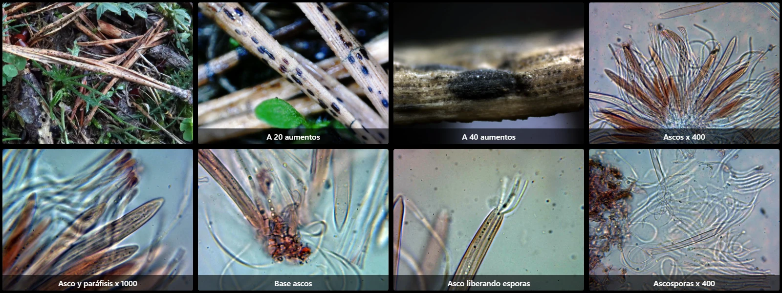
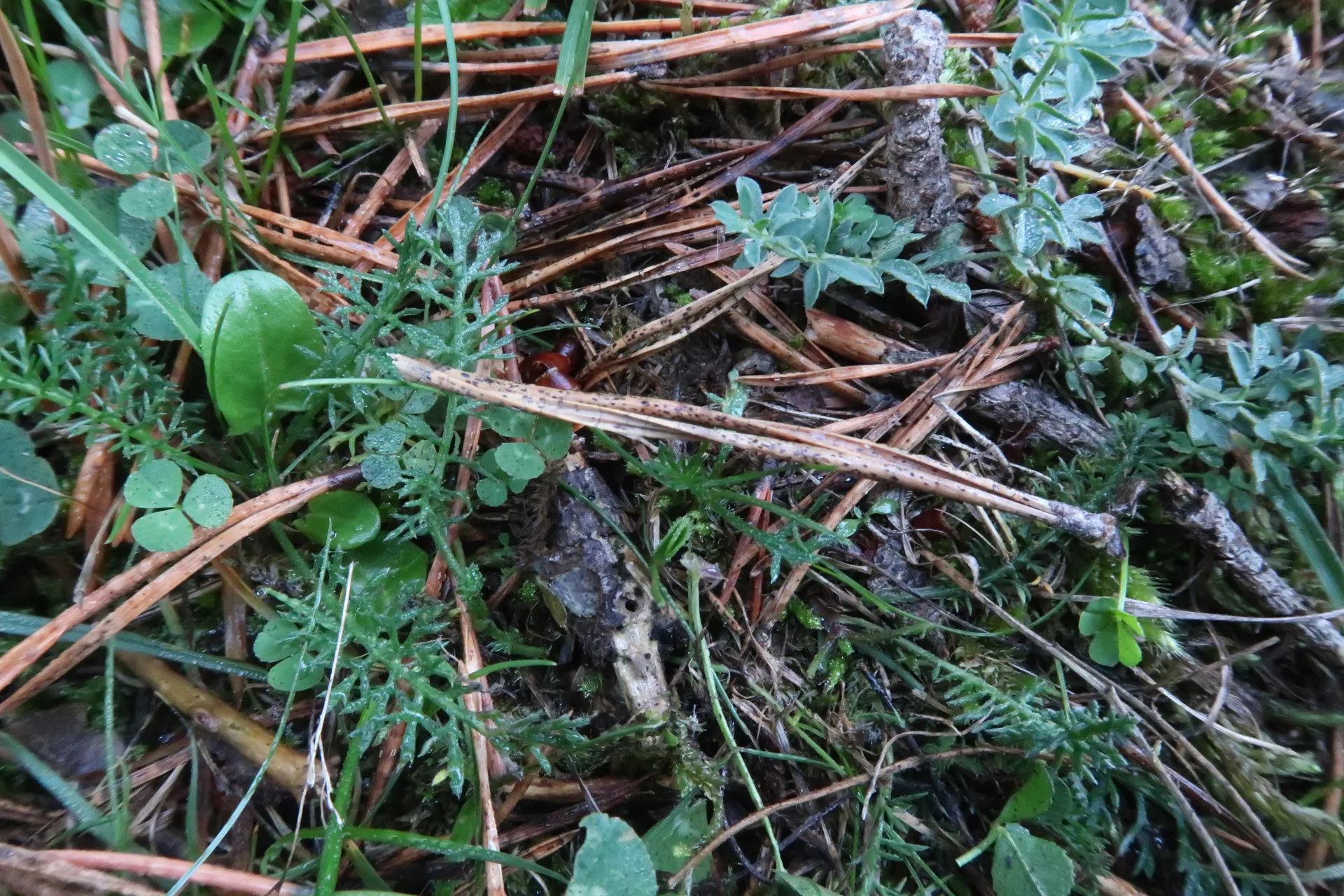
Substrate: Developing on decaying pine needles, particularly Pinus sylvestris.
Ascomata: Hysterothecia blackish, elliptical, 0.5-1.2 mm long and 0.5-0.8 mm wide, with a longitudinal slit extending over more than three quarters of the ascoma length.
Peridium: Composed of thick-walled cells of textura prismatica with dense blackish-brown intracellular pigment.
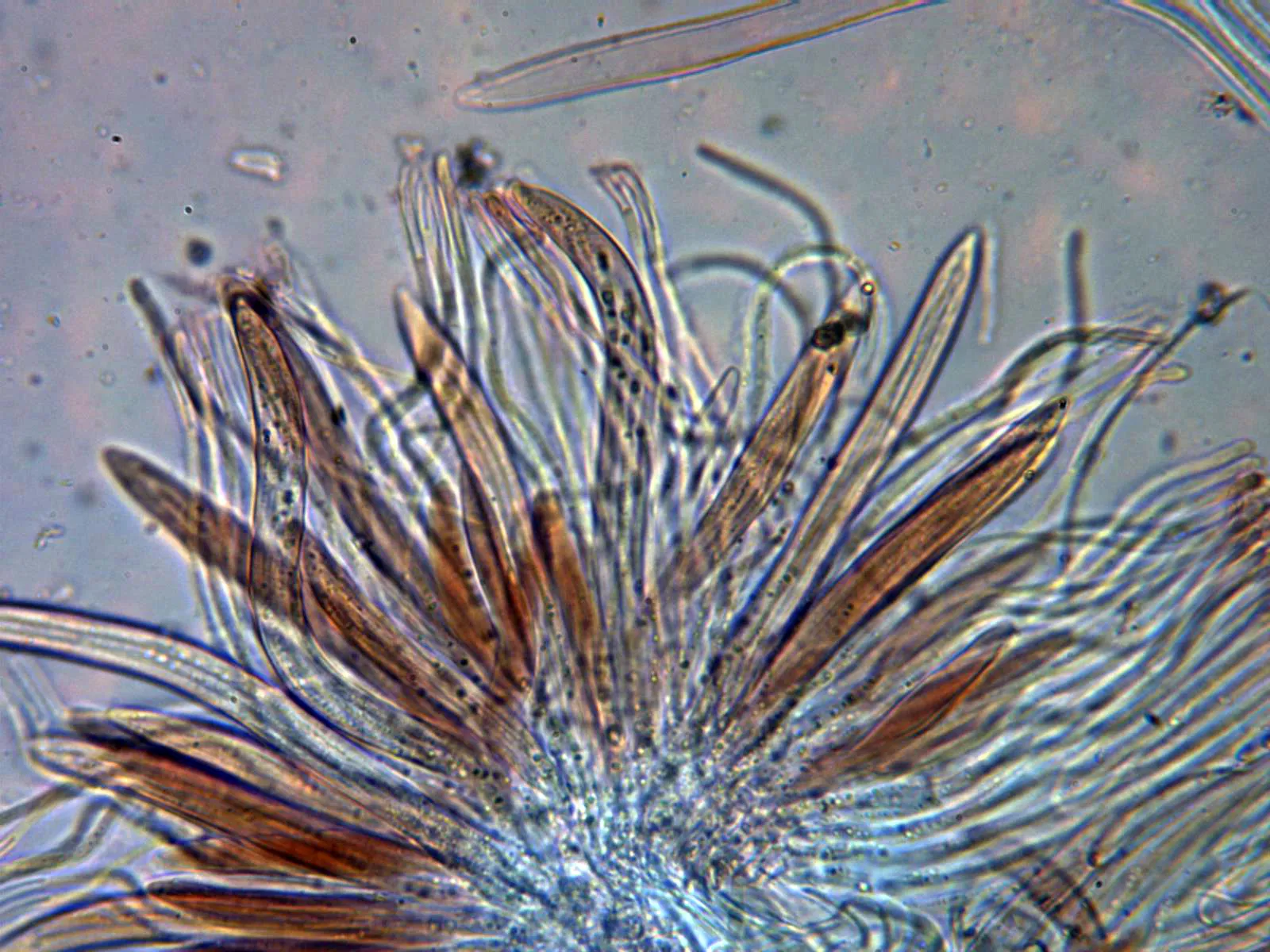
Asci: Cylindrical to slightly clavate, unitunicate, 120-150 x 10-13 µm, 8-spored, spores arranged in parallel fascicles within the ascus, without basal croziers, inamyloid in Melzer's.
Paraphyses: Filiform, smooth and thin-walled, unbranched, non-septate, 1-2 µm wide, apex not swollen, not projecting beyond asci.
Ascospores: Filiform, smooth-walled with pointed ends, difficult to measure, reported in literature as 80-110 x 1.5-2 µm, aseptate.
Ascomata: Hysterothecia blackish, elliptical, 0.5-1.2 mm long and 0.5-0.8 mm wide, with a longitudinal slit extending over more than three quarters of the ascoma length.
Peridium: Composed of thick-walled cells of textura prismatica with dense blackish-brown intracellular pigment.
Asci: Cylindrical to slightly clavate, unitunicate, 120-150 x 10-13 µm, 8-spored, spores arranged in parallel fascicles within the ascus, without basal croziers, inamyloid in Melzer's.
Paraphyses: Filiform, smooth and thin-walled, unbranched, non-septate, 1-2 µm wide, apex not swollen, not projecting beyond asci.
Ascospores: Filiform, smooth-walled with pointed ends, difficult to measure, reported in literature as 80-110 x 1.5-2 µm, aseptate.
Geographic references
Map generated from global occurrence data from GBIF. GBIF.
Study measurements
Values in Microns (µm)Macroscopic gallery
1 / 6Lophodermium pinastri - 1 - IMG_3713
Microscopic gallery
1 / 7Lophodermium pinastri - 10 - Ascas x 400 2