


Hygrophorocybe nivea
(Velen.) Vizzini & Contu
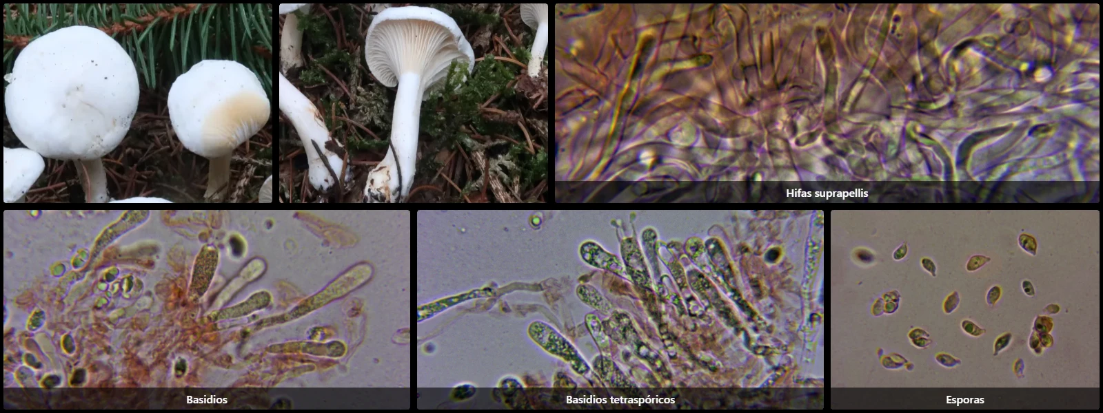
Study description
Formerly known as Clitocybe nivea, white with a pale cream spore print.
Pileipellis composed of filamentous, densely interwoven hyphae, thin- to moderately thick-walled, some branched, 3-6 µm wide, hyaline, septate with clamp connections at the septa.
Gill edge without differentiated elements from basidia or basidioles.
Basidia cylindric-clavate, 30-45 µm long and 6-10 µm wide, with basal clamps, mostly 4-spored.
Spores ellipsoidal to amygdaliform, smooth, hyaline, thin-walled, with a prominent apiculus at one end, vacuolar internal content, inamyloid in Melzer's reagent.
Geographic references
Map generated from global occurrence data from GBIF. GBIF.
Study measurements
Values in Microns (µm)Macroscopic gallery
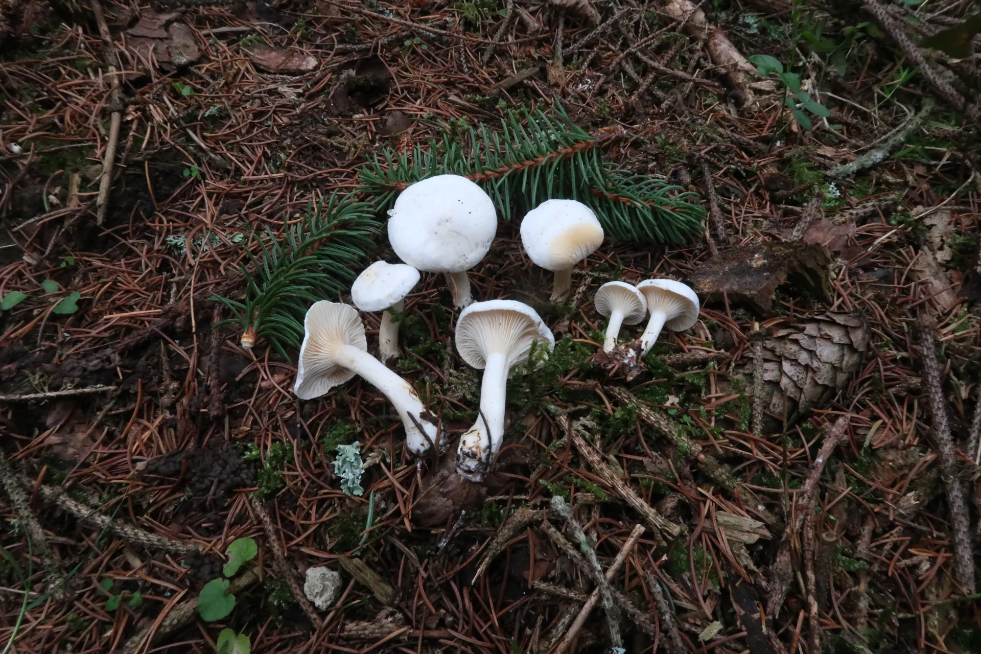
1 / 3Hygrophorocybe nivea - 1 - IMG_2176
Microscopic gallery
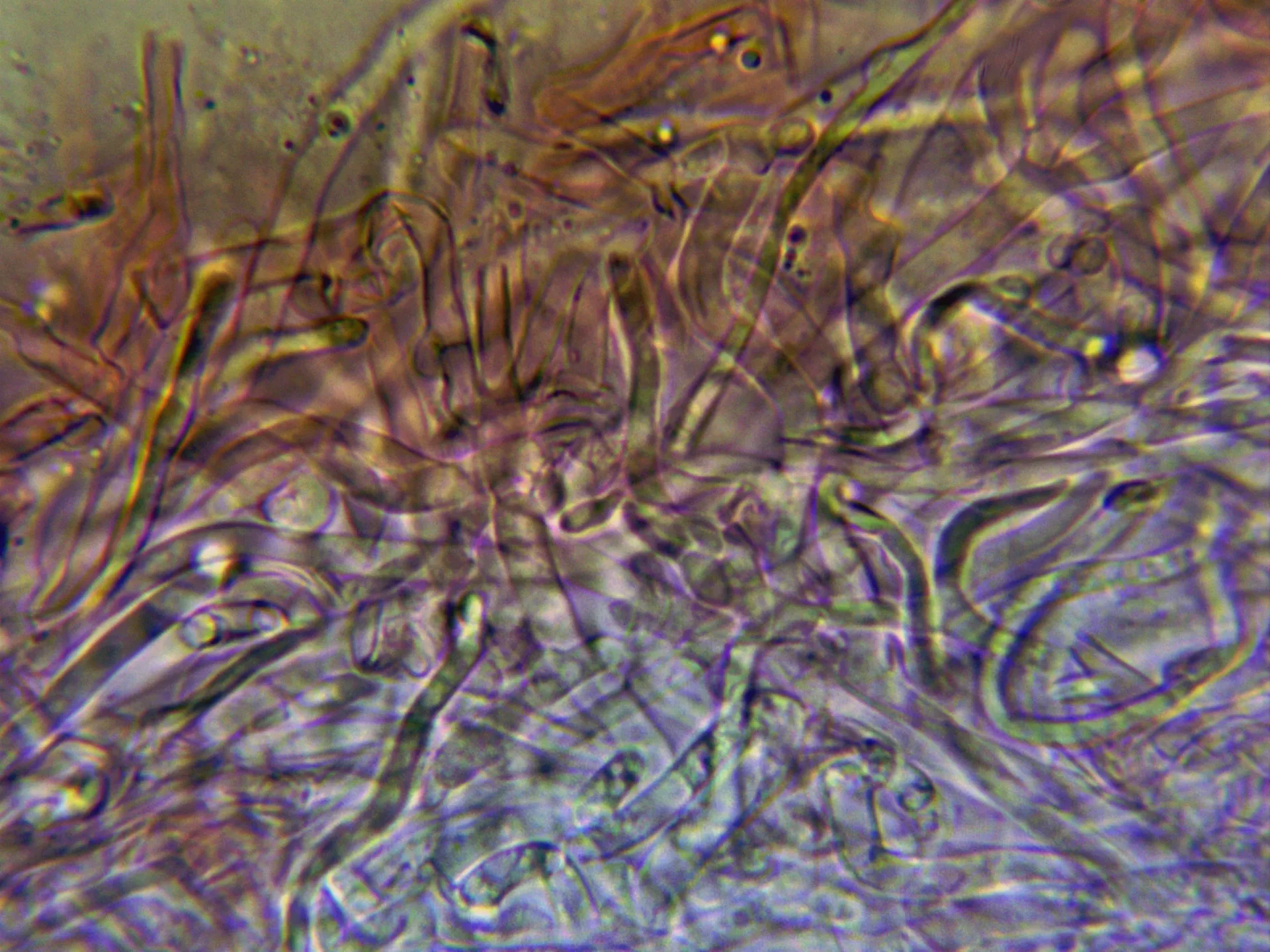
1 / 4Hygrophorocybe nivea - 6 - Suprapellis