


Ganoderma resinaceum
Boud.
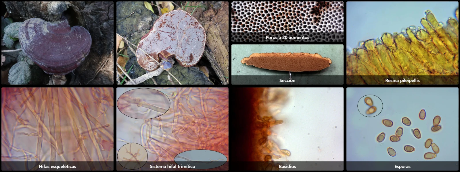
Study description
Trimitic hyphal system, with very thick-walled skeletal hyphae abundant in context (3 to 6 µm wide, unbranched, aseptate, with yellowish-brown pigment), thin-walled clamped generative hyphae (2 to 5 µm wide), and thick-walled, tortuous, branched binding hyphae up to 5 µm wide.
Hymenial cystidia and other sterile elements absent.
Basidia not observed, presumably 4-spored.
Spores ellipsoidal, with a truncate apex, double-walled, pale brown under the microscope with darker punctations, a large internal oil droplet, inamyloid in Melzer's reagent.
Geographic references
Map generated from global occurrence data from GBIF. GBIF.
Study measurements
Values in Microns (µm)Macroscopic gallery
1 / 10Ganoderma resinaceum - 1 -
Microscopic gallery
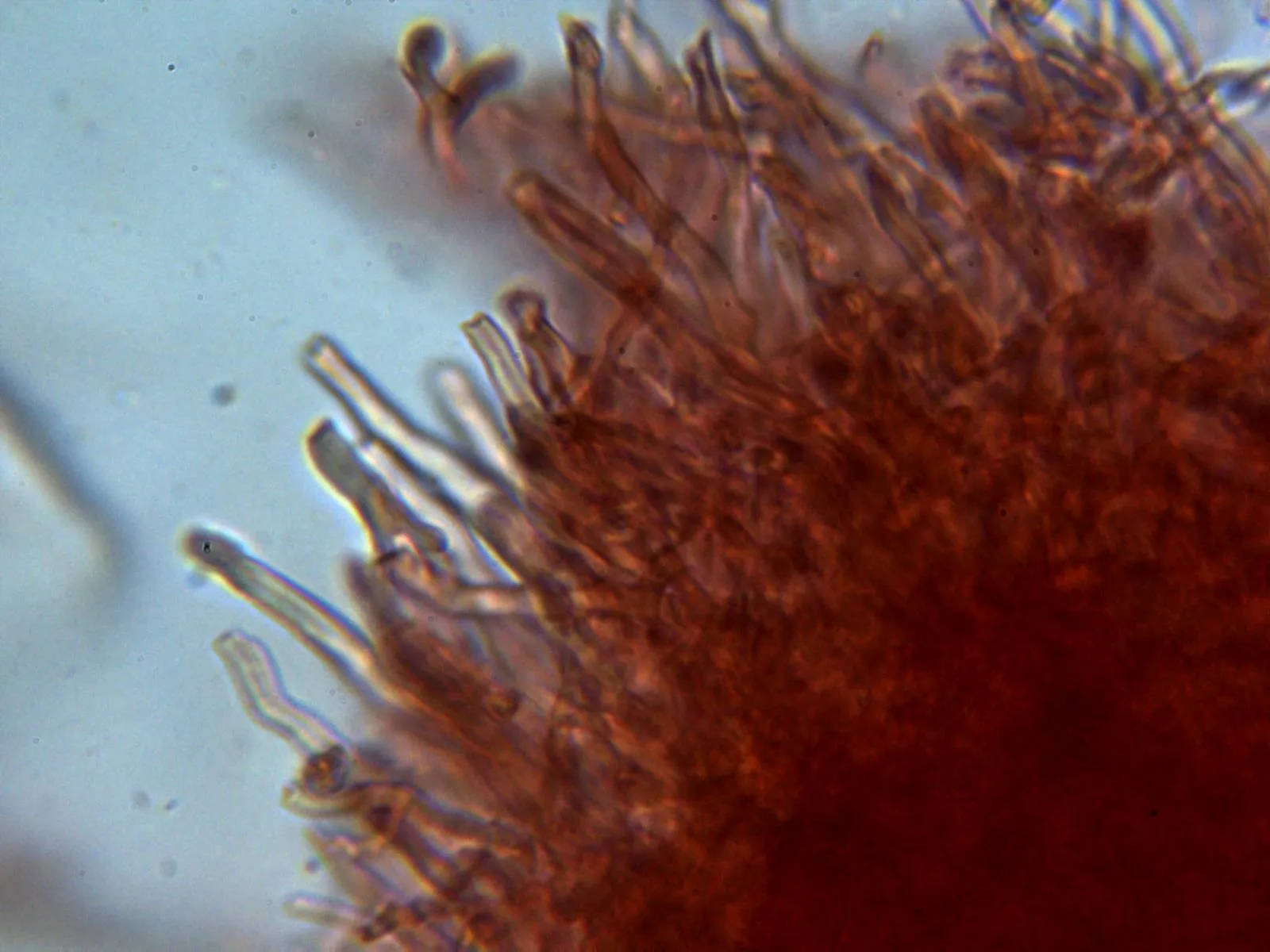
1 / 19Ganoderma resinaceum - 10 - Himenio 2