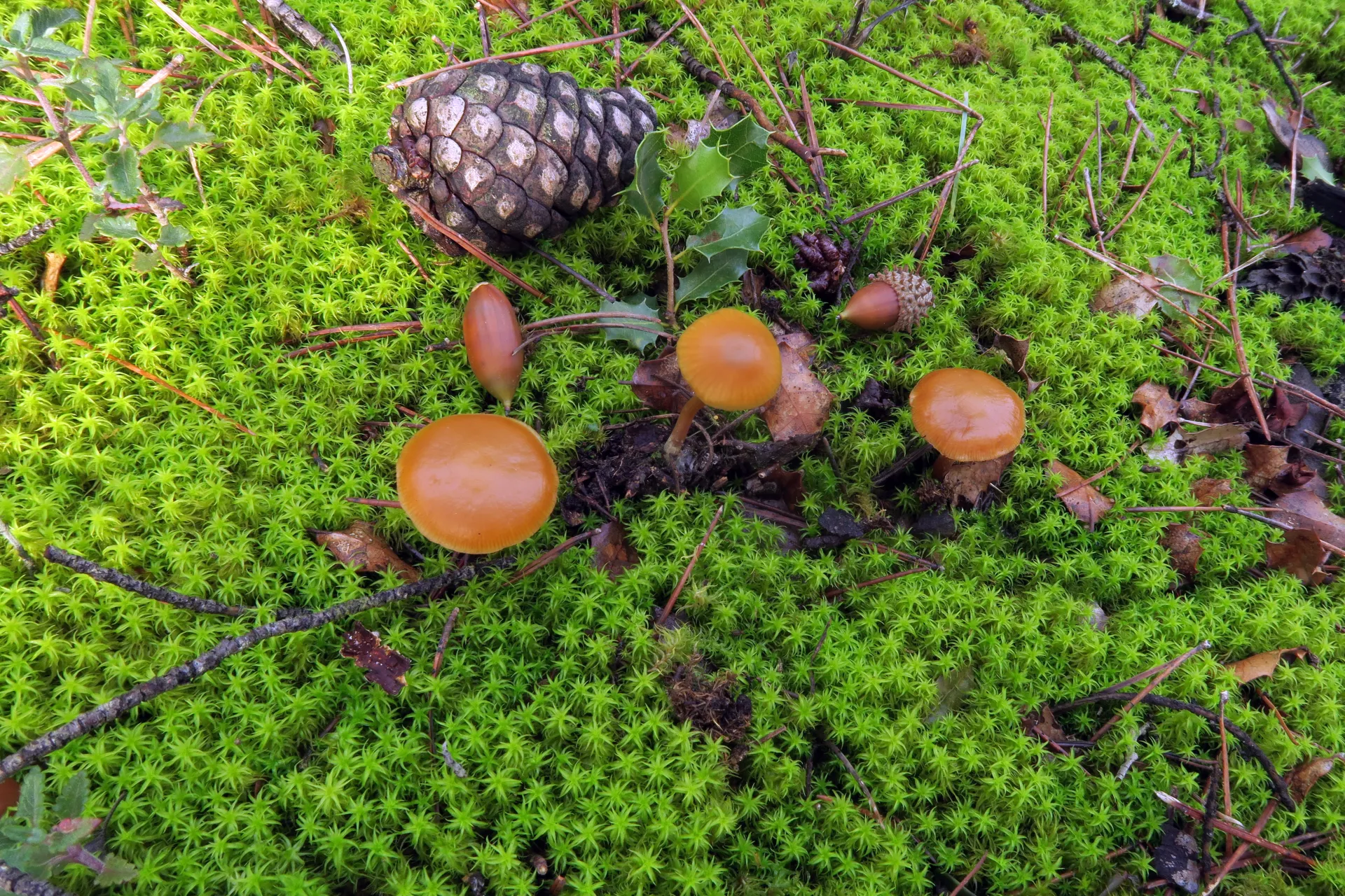
Galerina esteve-raventosii
Siquier, Olariaga, Salom & Høil.
Study description
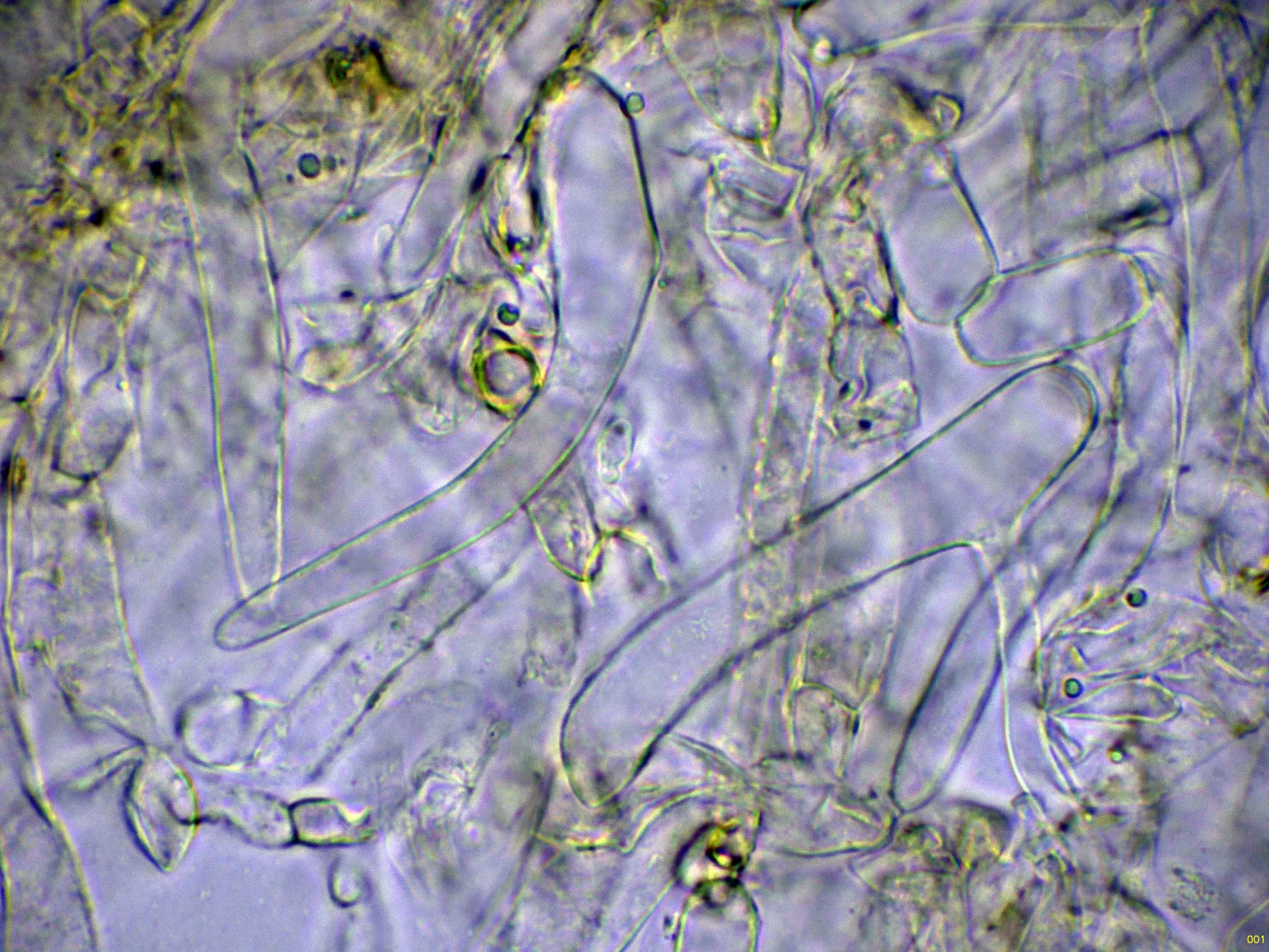
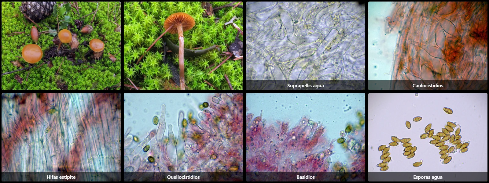
Filamentous cutis formed by interwoven hyphae, some branched, with faint yellowish intracellular pigment, constricted at septa.
Stipe hyphae arranged parallel, hyaline and clamped.
Caulocystidia present only on the upper stipe, generally fusiform, large (up to 70 µm long), some with a long neck and rounded apices.
Cheilocystidia very abundant, rendering the lamellar edge sterile, clustered in fascicles, up to 68 µm long by 11 µm wide, fusiform to lageniform, with a long sinuous neck and rounded tips.
Pleurocystidia scattered, of similar morphology to cheilocystidia.
Basidia cylindrical to very slightly ventricose, up to 34 µm long by 10 µm wide, with basal clamp connections, generally 4-spored, with occasional 2-spored basidia.
Spores ellipsoidal to subamygdaliform, smooth- and thick-walled, yellow under the microscope, with an eccentric apiculus at one end and a faint central germ pore at the opposite end, dextrinoid in Melzer's reagent.
Herbarium material available: JTP2699/25.
Geographic references
Map generated from global occurrence data from GBIF. GBIF.
Study measurements
Values in Microns (µm)Macroscopic gallery
Microscopic gallery