


Cortinarius cinnabarinus
Fr.
Study description
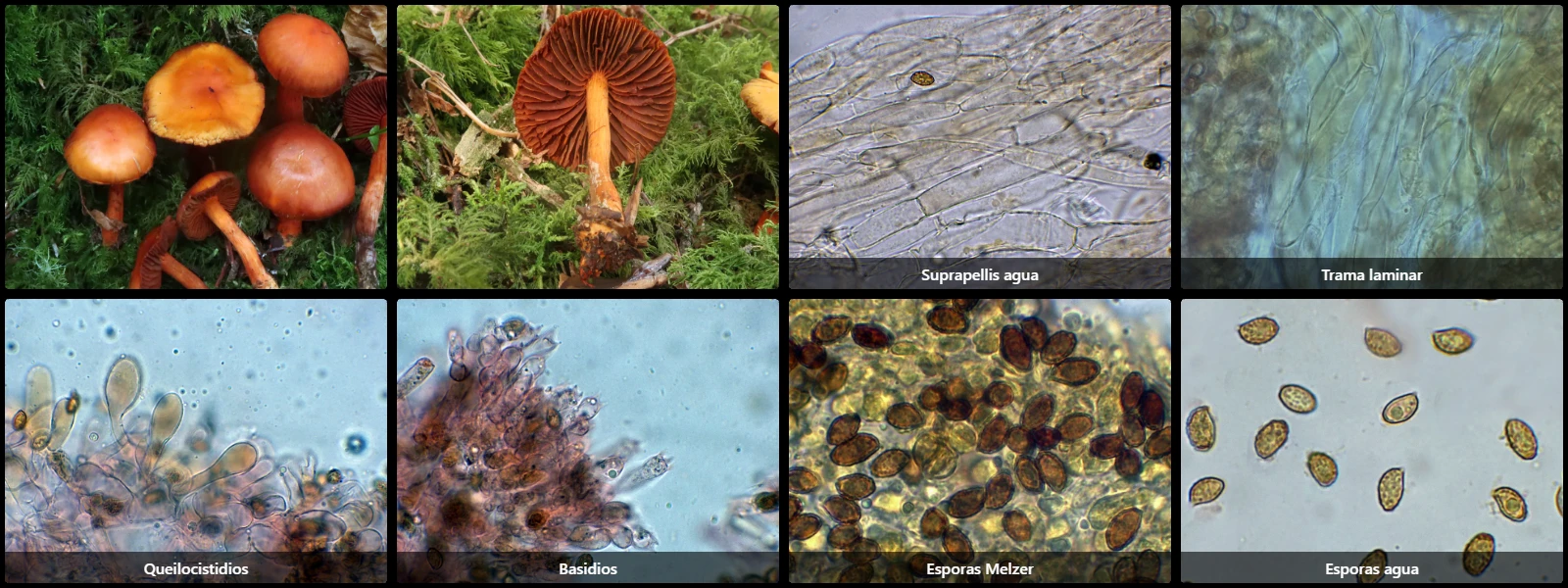
Suprapellis formed by a thin layer of narrow, parallel hyphae with ochraceous intracellular pigment.
Subpellis with a thick layer of very broad hyphae with short cells, also containing abundant intracellular pigment.
Cheilocystidia abundant on the lamellar edge, clavate, up to 28 µm long, strongly widened at the head, with abundant orange intracellular pigment.
Basidia cylindrical to slightly ventricose, up to 35 µm long, 4-spored.
Spores ellipsoidal to amygdaliform, thick-walled and verrucose, yellow under the microscope, with abundant darker warts, an eccentric apiculus, and dextrinoid reaction in Melzer's reagent.
Geographic references
Map generated from global occurrence data from GBIF. GBIF.
Study measurements
Values in Microns (µm)Macroscopic gallery
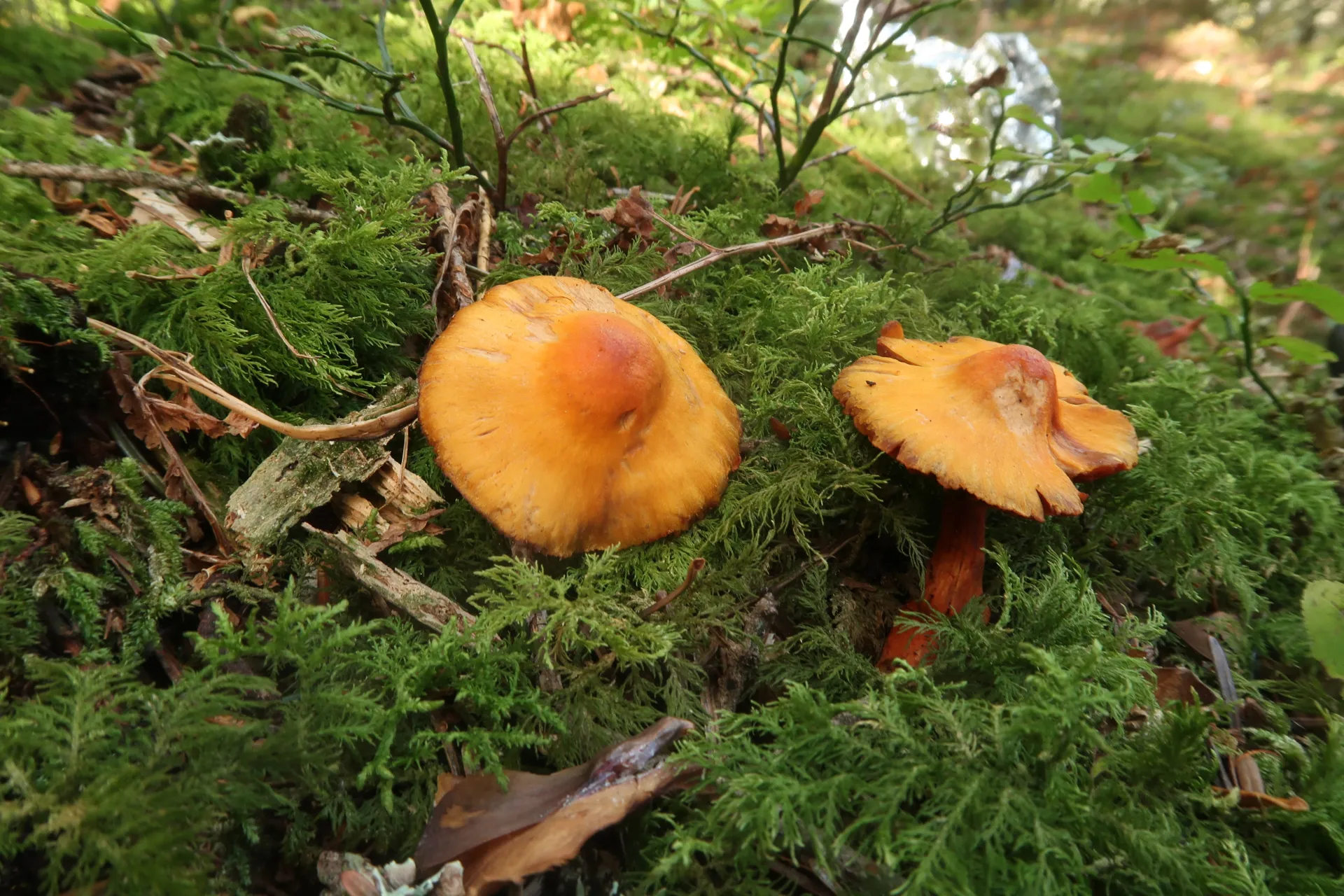
1 / 8Cortinarius cinnabarinus - 1 - IMG_0263
Microscopic gallery
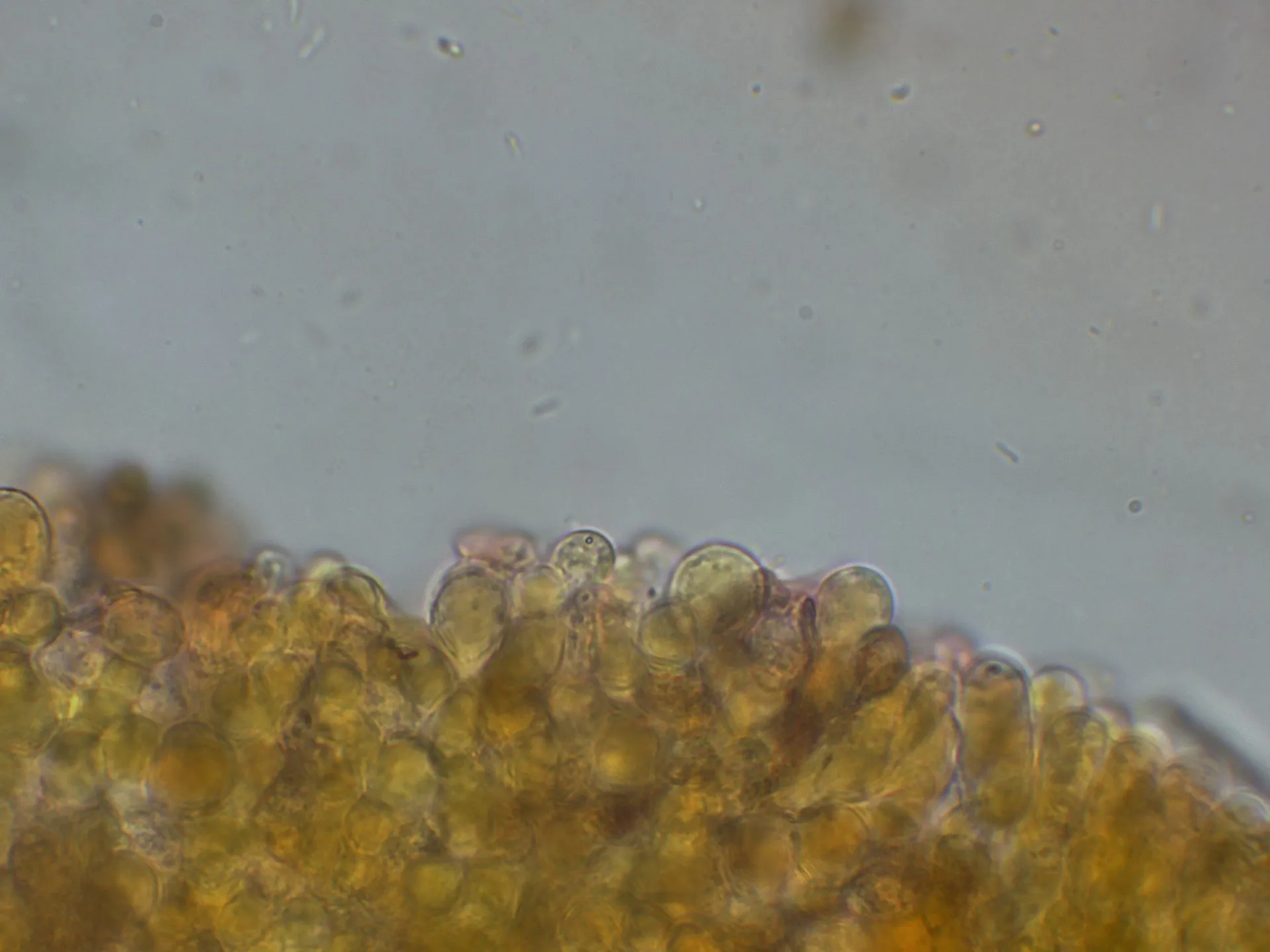
1 / 24Cortinarius cinnabarinus - 10 - Arista laminar-003
Genetic / Phylogenetic Study
Phylogenetic Tree
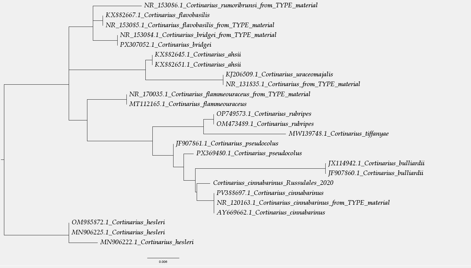
Arbol filogenético.png
Sequences and Data
Sequencing (Chromatogram)
Select a file to visualize the chromatogram