


Cortinarius casimirii
(Velen.) Huijsman
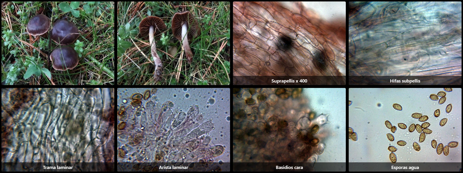
Study description
Pileipellis composed of two well-differentiated layers: an upper thin layer of narrow, parallel, heavily pigmented hyphae with clamped septa, and underneath a thicker layer of hyaline, parallel, much broader hyphae with short cells, even subglobose.
Lamellar edge homomorphic, without differentiated elements from basidia or basidioles.
Basidia clavate, up to 40 µm long, 4-spored.
Spores ellipsoidal to amygdaliform, yellowish-brown under the microscope, with an inconspicuous eccentric apiculus and hilar depression, moderately to strongly verrucose, with a marked dextrinoid reaction in Melzer's reagent.
Geographic references
Map generated from global occurrence data from GBIF. GBIF.
Study measurements
Values in Microns (µm)Macroscopic gallery
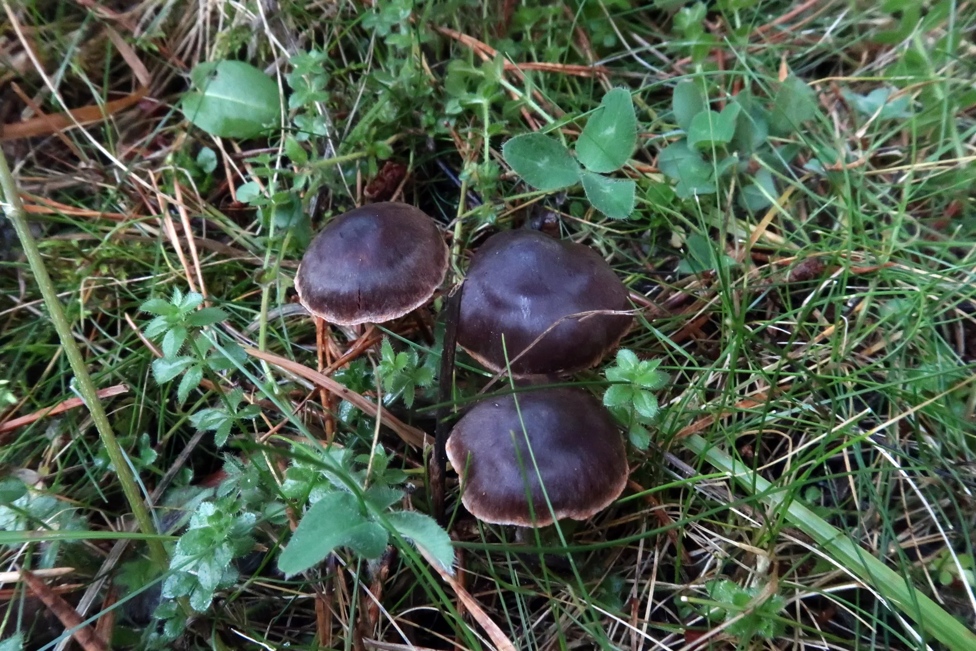
1 / 8Cortinarius casimirii - 1 - IMG_5722
Microscopic gallery
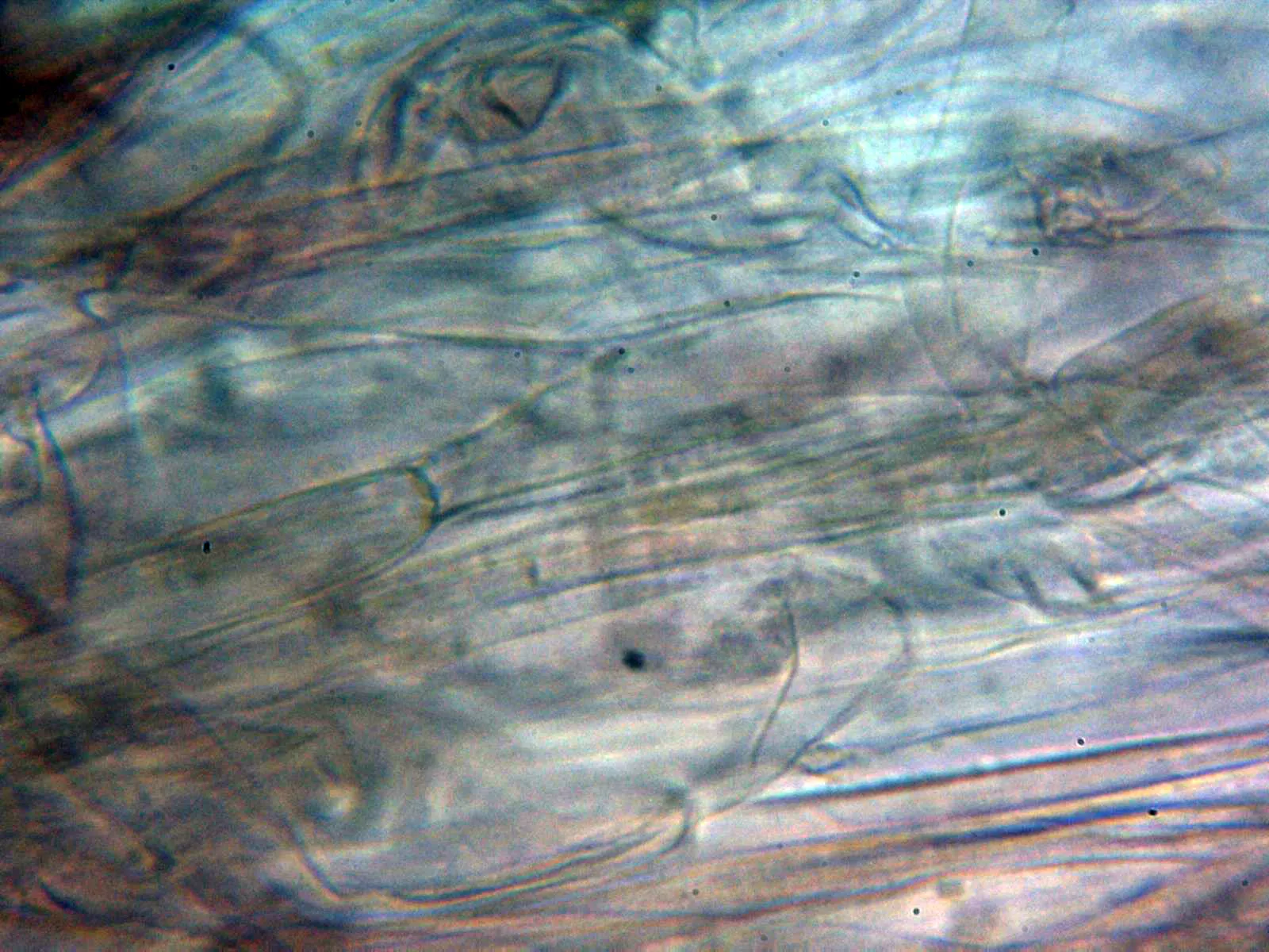
1 / 28Cortinarius casimirii - 10 - Hifas subpellis