


Coniophora fusispora
(Cooke & Ellis) Cooke
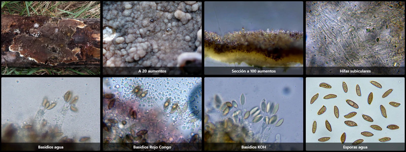
Study description
Subicular hyphae virtually absent and very difficult to observe, practically hyaline, with some swellings and without observed clamp connections.
Hymenium with basidia only, without differentiated cystidia.
Basidia cylindrical, very elongated, without swellings, without basal clamp connections, with faint internal content that decolorizes in KOH solution, 4-spored.
Spores pigmented, fusiform to navicular, with granular content, thick-walled, with a prominent eccentric apiculus at one end, virtually without reaction in Melzer's reagent (inamyloid), moderately decolorizing upon KOH application.
Herbarium material available: JTP2218/24.
Geographic references
Map generated from global occurrence data from GBIF. GBIF.
Study measurements
Values in Microns (µm)Macroscopic gallery
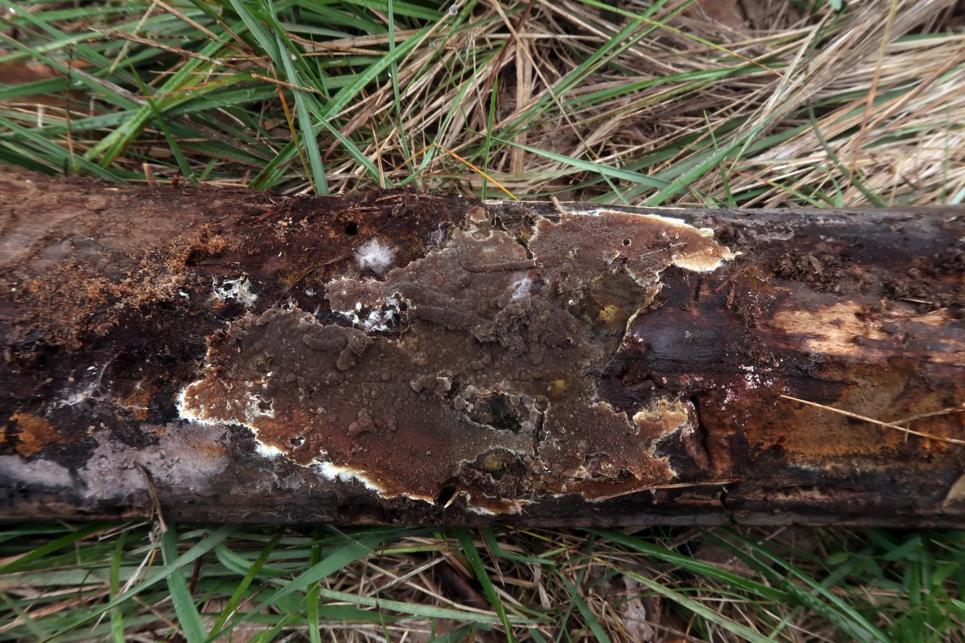
1 / 11Coniophora fusispora - 1 - IMG_0034
Microscopic gallery
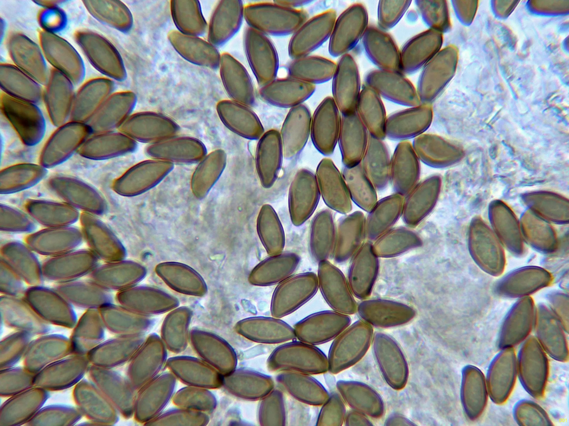
1 / 23Coniophora fusispora - 11 - Esporas KOH-001