


Calonarius alcalinophilus
(Rob.Henry) Niskanen & Liimat.
Study description
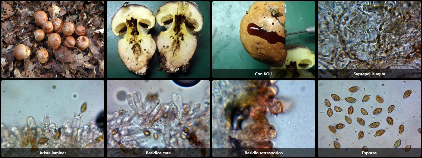
Suprapellis composed of filamentous, interwoven hyphae, widely separated by gelatinization, rather slender (2 to 5 µm wide), with abundant ochraceous intracellular pigment and clamped.
Lamellar edge without clearly differentiated elements from basidia, but with basidioliform terminal cells smaller than the basidia.
Basidia clavate and 4-spored, with basal clamp connections, up to 30 µm long; some basidia observed with ochraceous granular content.
Spores amygdaliform to citriform, rugose-walled, yellow under the microscope, with coarse darker warts and a prominent lateral apiculus.
Geographic references
Map generated from global occurrence data from GBIF. GBIF.
Study measurements
Values in Microns (µm)Macroscopic gallery
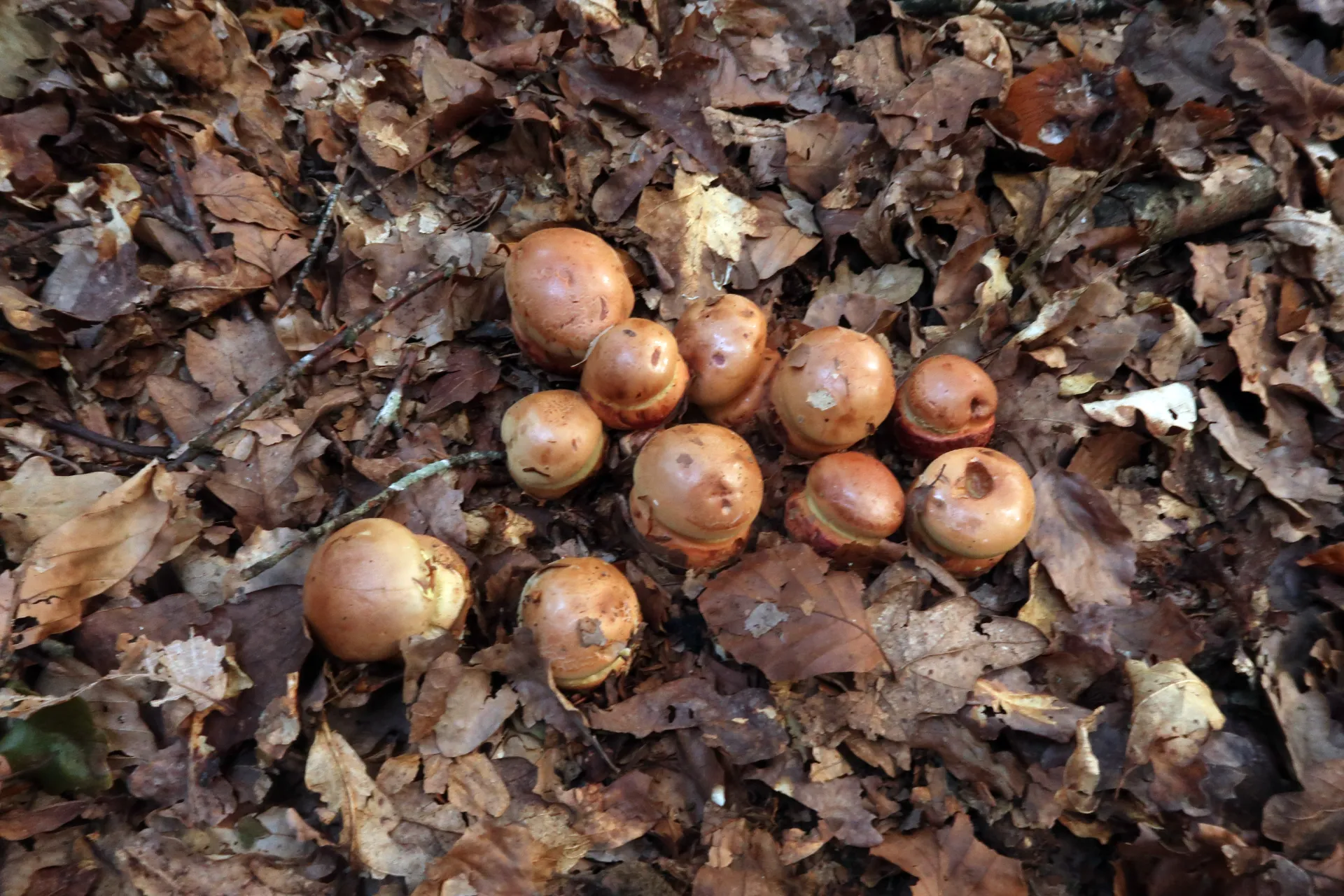
1 / 8Calonarius alcalinophilus - 1 - IMG_7849
Microscopic gallery
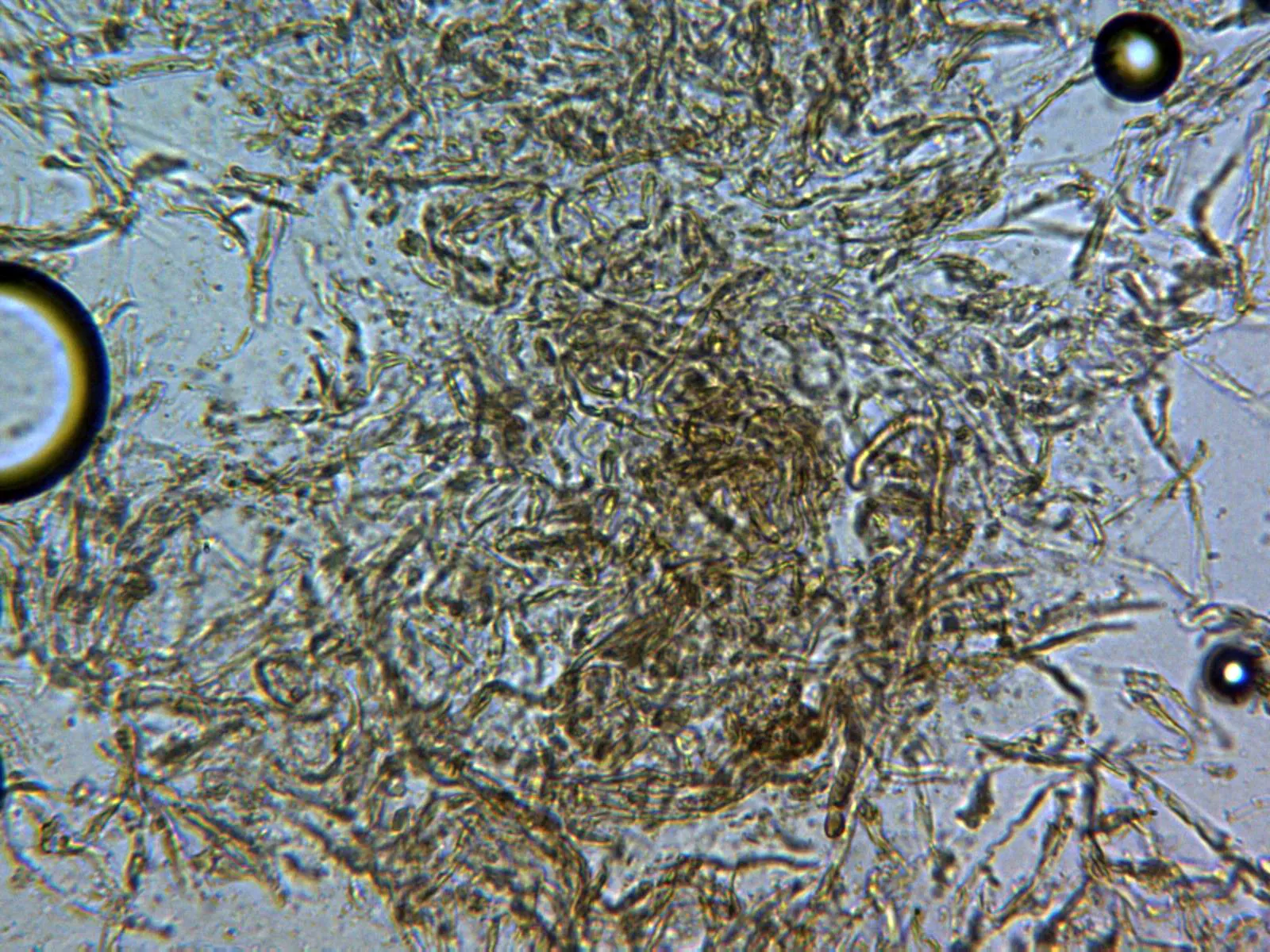
1 / 11Calonarius alcalinophilus - 11 - Suprapellis agua x 400